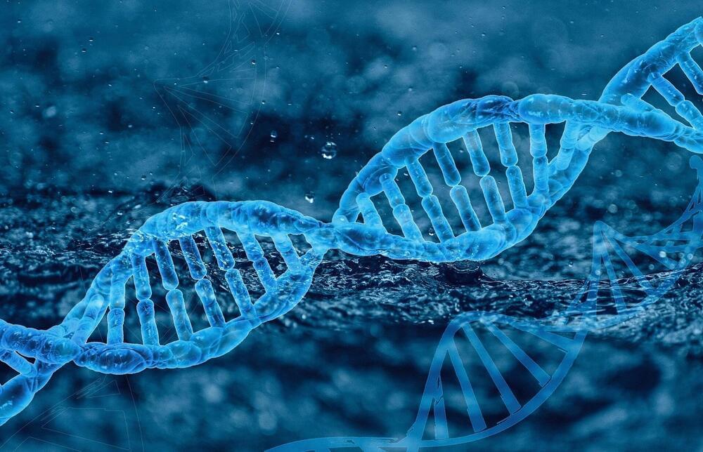
The structure of how DNA is stored in archaea makes a significant difference to how quickly it evolves, according to a new study by Indiana University researchers.
The study, led by molecular biologist Stephen Bell, Distinguished Professor and chair of the College of Arts and Sciences’ Department of Molecular and Cellular Biochemistry at Indiana University (IU) Bloomington, was recently published in Nature Microbiology. Its findings have the potential to impact research on the treatment of genetic diseases such as cancer.
“The most exciting thing we revealed is the idea that the shape of a DNA molecule can affect its ability to change,” Bell said. “In the early 20th century, modernist architecture had the idea that the form of a building should follow its function. But what we’re seeing in these organisms is that over time, form is actually affecting evolution. How DNA is structured can change it, creating an evolutionary feedback loop.”
(https://hscrb.harvard.edu/labs/whited-lab/) is an Assistant Professor of Stem Cell and Regenerative Biology at Harvard University where her lab focuses on limb regeneration in axolotl salamanders and where they develop tools to manipulate gene expression during limb regeneration, and explore signaling events following wound healing that initiate the regenerative process.
Dr. Whited earned a B.A. in Philosophy and a B.S. in Biological Sciences from the University of Missouri, and obtained her Ph.D. in Biology from MIT, where she studied in Dr. Paul Garrity’s laboratory.
Dr. Whited’s thesis focused on molecular mechanisms controlling the development and maintenance of cellular architectures in the Drosophila nervous system. During this work, Dr. Whited became interested in processes that may be required long after initial developmental events to ensure cells do not revert to immature behaviors, as well as processes that provoke such events in response to injury. She worked in the laboratory of Dr. Cliff Tabin (Harvard Medical School Department of Genetics) as a postdoc studying total limb regeneration in axolotl salamanders. During this time, Whited developed several molecular tools that can be used to interrogate regenerating axolotl limbs, which is one of the core focuses of her lab today.
Dr. Whited is also Co-Founder of Matice Biosciences, a company leveraging regenerative biology for the next generation of skincare and consumer scar products.
David Sinclair shares another side of himself. Compassion for all people. He wants to make sure that longevity technologies are available for all people, not just for the super wealthy and their pets. He also speaks of emerging elderly populations who can live well up until death rather than suffering for so long, and instead start new careers and hobbies.
Researchers have restored vision in animal by resetting some of the thousands of chemical marks that accumulate on DNA as cells age. The work, by Dr David Sinclair Lab, published in Nature Dec 2020, suggests a new approach to reversing age-related decline, by reprogramming some cells to a ‘younger’ state in which they are better able to repair or replace damaged tissue.
David A. Sinclair, Ph.D. A.O. is a tenured Professor in the Genetics Department at the Blavatnik Institute, Harvard Medical School, Boston & Co-Director of the Paul F. Glenn Center for Biology of Aging Research, honorary Professor at the University of Sydney, and co-founder of the journal Aging. He obtained a BS and a Ph.D. at UNSW, worked as a postdoctoral researcher at M.I.T., was hired at Harvard Medical School in 1999 as an Assistant Professor, and promoted to tenured Professor in 2008. His book Lifespan: Why We Age and Why We Don’t Have To, a NYT bestseller, is published in more than 20 languages.
Dr. Sinclair is an inventor on more than 50 patents, 170 papers, an h-index of 103 & cited 73,000+ times. His more than 40 awards include an Excellence in Teaching Award, Harvard, AFAR Fellowship, the Ellison Medical Foundation Scholarships, Genzyme Outstanding Achievement Award, Telluride Technology Award, Innovator of the Year, MERIT Award, Nathan Shock Award, Denham Harman Award, ASMR Medal, Advance Global Australian, Pioneer Award, TIME100’s most influential people, TIME magazine’s Heathcare 50, Irving Wright Award, AFAR, and is an Officer of the Order of Australia (AO).
He cofounded Sirtris Pharma (Cambridge; NASDAQ: SIRT, bought by GSK), Genocea (Cambridge, MA; NASDAQ: GNCA); Ovascience (NASDAQ: OVAS), Cohbar (Menlo Park NASDAQ: CWBR)), MetroBiotech, ArcBio, Liberty Biosecurity, Galilei, Immetas, EdenRoc Sciences and affiliates, and Life Biosciences and affiliates.
Ergothioneine exhibits longevity-extension effect in Drosophila melanogaster via regulation of cholinergic neurotransmission, tyrosine metabolism, and fatty acid oxidation. https://pubmed.ncbi.nlm.nih.gov/34877949/
17-a-estradiol late in life extends lifespan in aging UM-HET3 male mice; nicotinamide riboside and three other drugs do not affect lifespan in either sex. https://pubmed.ncbi.nlm.nih.gov/33788371/
Metagenomic and metabolomic remodeling in nonagenarians and centenarians and its association with genetic and socioeconomic factors.
By making remarkable breakthroughs in a number of fields, unlocking new approaches to science, and accelerating the pace of science and innovation.
In 2020, Google’s AI team DeepMind announced that its algorithm, AlphaFold, had solved the protein-folding problem. At first, this stunning breakthrough was met with excitement from most, with scientists always ready to test a new tool, and amusement by some. After all, wasn’t this the same company whose algorithm AlphaGo had defeated the world champion in the Chinese strategy game Go, just a few years before? Mastering a game more complex than chess, difficult as that is, felt trivial compared to the protein-folding problem. But AlphaFold proved its scientific mettle by sweeping an annual competition in which teams of biologists guess the structure of proteins based only on their genetic code. The algorithm far outpaced its human rivals, posting scores that predicted the final shape within an angstrom, the width of a single atom. Soon after, AlphaFold passed its first real-world test by correctly predicting the shape of the SARS-CoV-2 ‘spike’ protein, the virus’ conspicuous membrane receptor that is targeted by vaccines.
The success of AlphaFold soon became impossible to ignore, and scientists began trying out the algorithm in their labs. By 2021 Science magazine crowned an open-source version of AlphaFold the “Method of the Year.” Biochemist and Editor-in-Chief H. Holden Thorp of the journal Sciencewrote in an editorial, “The breakthrough in protein-folding is one of the greatest ever in terms of both the scientific achievement and the enabling of future research.” Today, AlphaFold’s predictions are so accurate that the protein-folding problem is considered solved after more than 70 years of searching. And while the protein-folding problem may be the highest profile achievement of AI in science to date, artificial intelligence is quietly making discoveries in a number of scientific fields.
By turbocharging the discovery process and providing scientists with new investigative tools, AI is also transforming how science is done. The technology upgrades research mainstays like microscopes and genome sequencers 0, adding new technical capacities to the instruments and making them more powerful. AI-powered drug design and gravity wave detectors offer scientists new tools to probe and control the natural world. Off the lab bench, AI can also deploy advanced simulation capabilities and reasoning systems to develop real-world models and test hypotheses using them. With manifold impacts stretching the length of the scientific method, AI is ushering in a scientific revolution through groundbreaking discoveries, novel techniques and augmented tools, and automated methods that advance the speed and accuracy of the scientific process.
Cells not replaced, but old cells that are still there are rejuvenated.
Dr David Sinclair explains the mechanism behind how to reprogramm the old cells rejuvenate to be young again. He also clarify the process is based on cell autonomous effect and does not involve or rely on any stem cells in this clip.
David Sinclair is a professor in the Department of Genetics and co-director of the Paul F. Glenn Center for the Biology of Aging at Harvard Medical School, where he and his colleagues study sirtuins—protein-modifying enzymes that respond to changing NAD+ levels and to caloric restriction—as well as chromatin, energy metabolism, mitochondria, learning and memory, neurodegeneration, cancer, and cellular reprogramming.
Dr David Sinclair has suggested that aging is a disease—and that we may soon have the tools to put it into remission—and he has called for greater international attention to the social, economic and political and benefits of a world in which billions of people can live much longer and much healthier lives.
Dr David Sinclair is the co-founder of several biotechnology companies (Life Biosciences, Sirtris, Genocea, Cohbar, MetroBiotech, ArcBio, Liberty Biosecurity) and is on the boards of several others.
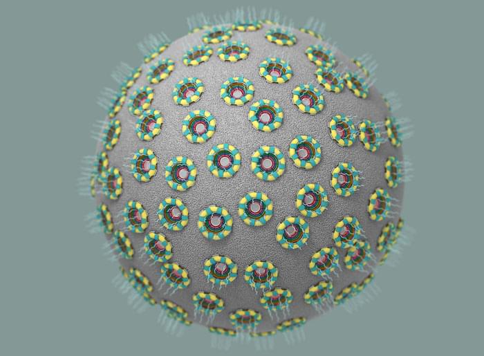
Whatever you are doing, whether it is driving a car, going for a jog, or even at your laziest, eating chips and watching TV on the couch, there is an entire suite of molecular machinery inside each of your cells hard at work. That machinery, far too small to see with the naked eye or even with many microscopes, creates energy for the cell, manufactures its proteins, makes copies of its DNA, and much more.
Among those pieces of machinery, and one of the most complex, is something known as the nuclear pore complex (NPC). The NPC, which is made of more than 1,000 individual proteins, is an incredibly discriminating gatekeeper for the cell’s nucleus, the membrane-bound region inside a cell that holds that cell’s genetic material. Anything going in or out of the nucleus has to pass through the NPC on its way.
Nuclear pores stud the surface of the cell’s nucleus, controlling what flows in and out of it. (Image: Valerie Altounian)
“When the Human Genome Project began in 1990, it had a projected budget of $3 billion. […] Now, one company claims to have achieved the major milestone of whole genome sequencing for just $100.”
Ultima Genomics, a biotech company based in California, has emerged from stealth mode with a new high-throughput, low-cost sequencing platform that it claims can deliver a $100 genome.
When the Human Genome Project began in 1990, it had a projected budget of $3 billion. Some researchers believed it would take centuries to map all 20,000+ genes and to determine the sequence of chemical base pairs making up DNA, though in the end it took 13 years. Since then, genome sequencing has undergone technology and cost improvements at a rate faster than Moore’s Law (a long-term trend in the computer industry that involves a doubling of performance every two years). What used to require billions of dollars and many years of work is now several orders of magnitude cheaper and possible in a matter of hours.
Companies like 23andMe and Ancestry.com have been offering DNA test kits at the consumer level. These can generate reports relating to a customer’s ancestry and genetic predispositions to health-related issues. While most people have opted for tests based on partial (i.e. incomplete) sequencing, the costs are now becoming so low that whole genome sequencing may soon be affordable. Veritas Genetics made headlines in 2016 by breaking the $1,000 barrier and in 2021 the price fell to $562.
Not that Thier should be more animals raised for meat.
But beefalo does have its opponents.
“We just don’t think there should be beefalo,” said Martha McFarland, farmland viability coordinator for the advocacy group Practical Farmers of Iowa. She also raises cattle and bison, but said she would never mix the two.
“Nature did just fine producing bison. It’s an excellent animal that also is good to eat, and mixing it with cows is not necessary and weakens the genetic line of the bison.”
Having multiple conditions that affect the heart are linked to a greater risk of dementia than having high genetic risk, according to a largescale new study.
Led by Oxford University and the University of Exeter, the study is among the largest ever to examine the link between several heart-related conditions and dementia, and one of the few to look at the complex issue of multiple health conditions.
Published in The Lancet Healthy Longevity, the paper looked at data from more than 200,000 people, aged 60 or above, and of European ancestry in UK Biobank. The international research team identified those who had been diagnosed with the cardiometabolic conditions diabetes, stroke, or a heart attack, or any combination of the three, and those who went on to develop dementia.