Toggle light / dark theme

The great powerful guppy can essentially evolve 10 million times faster than usual. Which could lead to humans evolving faster too leading to a biological singularity.


Although natural selection is often viewed as a slow pruning process, a dramatic new field study suggests it can sometimes shape a population as fast as a chain saw can rip through a sapling. Scientists have found that guppies moved to a predator-free environment adapted to it in a mere 4 years—a rate of change some 10,000 to 10 million times faster than the average rates gleaned from the fossil record. Some experts argue that the 11-year study, described in today’s issue of Science,* may even shed light on evolutionary patterns that occur over eons.

A team led by evolutionary biologist David Reznick of the University of California, Riverside, scooped guppies from a waterfall pool brimming with predators in Trinidad’s Aripo River, then released them in a tributary where only one enemy species lurked. In as little as 4 years, male guppies in the predator-free tributary were already detectably larger and older at maturity when compared with the control population; 7 years later females were too. Guppies in the safer waters also lived longer and had fewer and bigger offspring.

The team next determined the rate of evolution for these genetic changes, using a unit called the darwin, or the proportional amount of change over time. The guppies evolved at a rate between 3700 and 45,000 darwins. For comparison, artificial-selection experiments on mice show rates of up to 200,000 darwins—while most rates measured in the fossil record are only 0.1 to 1.0 darwin. “It’s further proof that evolution can be very, very fast and dynamic,” says Philip Gingerich, a paleontologist at the University of Michigan, Ann Arbor. “It can happen on a time scale that’s as short as one generation—from us to our kids.”

Many of the fundamental principles in biology and essentially all pathways regulating development were identified in so-called genetics screens. Originally pioneered in the fruit fly Drosophila and the nematode C. elegans, genetic screens involve inactivation of many genes one by one. By analyzing the consequences of gene loss, scientists can draw conclusions about its function. This way, for example, all genes required for formation of a brain can be identified.

Genetic screens can routinely be carried out in flies and worms. In humans, a wealth of knowledge exists about genetic disorders and the consequences of disease-relevant mutations, but their systematic analysis was impossible. Now, the Knoblich lab at IMBA has developed a groundbreaking technique allowing hundreds of to be analyzed in parallel in . They named the new technology CRISPR-LICHT and published their findings in the journal Science.

By using cerebral organoids, a 3D cell culture model for the human developed in Jürgen Knoblich’s group at IMBA, hundreds of mutations can now be analyzed for their role in the using CRISPR-LICHT.

While our circadian body clock dictates our preferred rhythm of sleep or wakefulness, a relatively new concept—the epigenetic clock—could inform us about how swiftly we age, and how prone we are to diseases of old age.

People age at different rates, with some individuals developing both characteristics and diseases related to aging earlier in life than others. Understanding more about this so-called ‘biological age’ could help us learn more about how we can prevent diseases associated with age, such as . Epigenetic markers control the extent to which genes are switched on and off across the different cell-types and tissues that make up a . Unlike our , these epigenetic marks change over time, and these changes can be used to accurately predict biological age from a DNA .

Now, scientists at the University of Exeter have developed a new specifically for the . As a result of using human tissue samples, the new clock is far more accurate than previous versions, that were based on blood samples or other tissues. The researchers hope that their new clock, published in Brain and funded by Alzheimer’s Society, will provide insight into how accelerated aging in the brain might be associated with brain diseases such as Alzheimer’s and other forms of dementia.

The coronavirus disease 2019 (COVID-19) pandemic does not affect everyone equally. While anyone can contract COVID-19, accumulating data suggest that older people or those with pre-existing comorbidities are far more likely to have severe complications or die from the disease. While researchers scramble to unravel the mechanisms of action underlying the disease’s wide-ranging effects, news that the disease hits older people hardest has been received without demur: it is widely accepted that to be old is to be fragile. Indeed, even in so-called normal times, everyone expects more things break as people age: bones, hearts, brains. In the context of the pandemic, being old is seen as just one more comorbidity.

It should not be.

We accept growing old and losing our vitality as an inevitability of life. To do so is to overlook the fact that ageing is, fundamentally, a plastic trait—influenced both by our genetic predispositions and many (controllable) environmental factors. Anecdotally we know this to be true: for some, being in their eighties means being confined to a wheelchair whereas for others, like Eileen Noble, who at 84 years old was the oldest runner in 2019’s London Marathon, it decidedly does not. The burgeoning field of biogerontology is now beginning to amass data in support of such observations. Single genetic mutations in evolutionarily conserved pathways across model organisms—ranging from fruit flies to mice—increase lifespan by up to 80%. Crucially, not only do these animals live longer, they also have a longer youthspan—the proportion of their lives in which they retain the trappings of youth such as peak mobility, immunity, and stress resilience.

Summary: A mutation in a gene associated with circadian rhythm extends the clock period, causing people to stay up late at night and sleep late in the mornings.

Source: UC Santa Cruz

A new study by researchers at UC Santa Cruz shows how a genetic mutation throws off the timing of the biological clock, causing a common sleep syndrome called delayed sleep phase disorder.

KENNEDY SPACE CENTER (FL), October 19, 2020 – The Center for the Advancement of Science in Space (CASIS) and the National Science Foundation (NSF) announced three flight projects that were selected as part of a joint solicitation focused on leveraging the International Space Station (ISS) U.S. National Laboratory to further knowledge in the fields of tissue engineering and mechanobiology. Through this collaboration, CASIS, manager of the ISS National Lab, will facilitate hardware implementation, in-orbit access, and astronaut crew time on the orbiting laboratory. NSF invested $1.2 million in the selected projects, which are seeking to advance fundamental science and engineering knowledge for the benefit of life on Earth.

This is the third collaborative research opportunity between CASIS and NSF focused on tissue engineering. Fundamental science is a major line of business for the ISS National Lab, and by conducting research in the persistent microgravity environment offered by the orbiting laboratory, NSF and the ISS National Lab will drive new advances that will bring value to our nation and spur future inquiries in low Earth orbit.

Microgravity affects organisms—from viruses and bacteria to humans, inducing changes such as altered gene expression and DNA regulation, changes in cellular function and physiology, and 3D aggregation of cells. Spaceflight is advancing research in the fields of pharmaceutical research, disease modeling, regenerative medicine, and many other areas within the life sciences. The selected projects will utilize the ISS National Lab and its unique environment to advance fundamental and transformative research that integrates engineering and life sciences.

“This is kind of a nice bookend to 16 years of research,” says Deisseroth, a neuroscientist and bioengineer at Stanford University. “It took years and years for us to sort out how to make it work.”

“The result is described this month in the journal Nature Biotechnology.”

“Optogenetics involves genetically engineering animal brains to express light-sensitive proteins—called opsins—in the membranes of neurons.”


Optogenetics can now control neural circuits at unprecedented depths within living brain tissue without surgery.

A team of researchers at Duke University have developed an imaging technology for tagging structures at a cellular level that overcomes the shortcomings of existing antibody-based techniques. Immunofluorescence imaging is a key part of the cell biologist’s toolbox, in which a fluorescent ‘flare’ attached to an antibody allows them to visualize the presence of specific target proteins in cell or tissue samples. The issue is that this specificity isn’t always 100 percent — sometimes the antibodies bind to other closely related proteins as well, making it difficult to interpret the results.

Duke’s cell biology chair Scott Soderling has led a team that developed Homology-independent Universal Genome Engineering (HiUGE), an innovation that uses gene-editing technology to rise above the shortcomings of traditional commercial antibodies for imaging.

“We had this idea that CRISPR could be a really amazing tool to address the pressing problem of trying to identify and label these hundreds of proteins,” said Soderling.

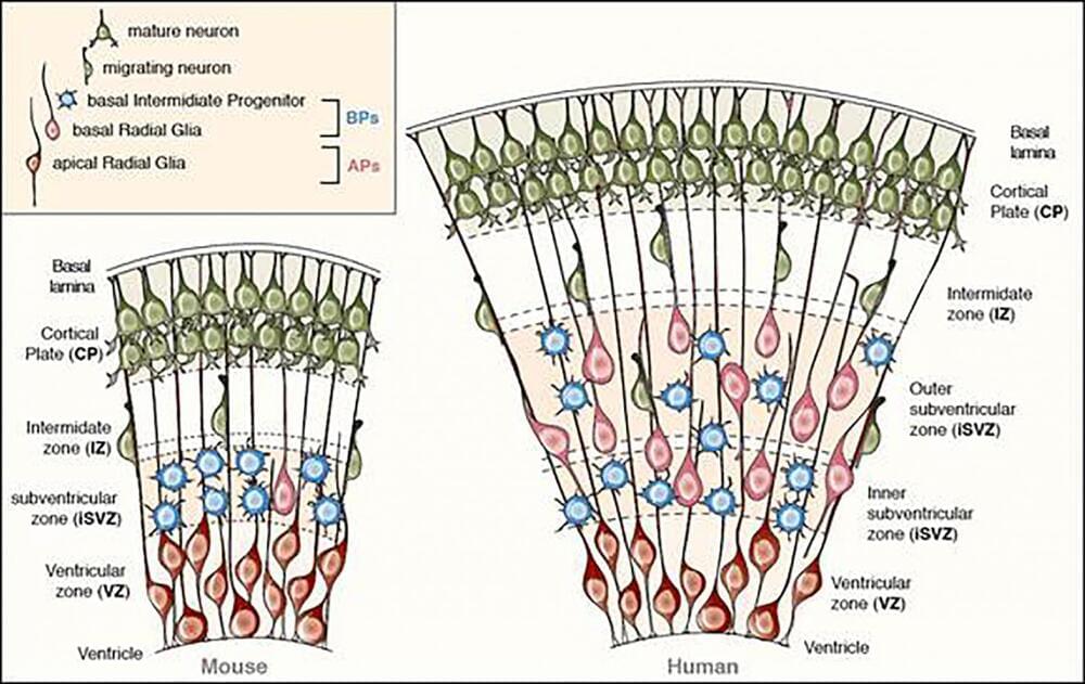
Summary: Study reveals a new role for serotonin in the development of the human neocortex. Serotonin acts cell-extrinsically as a growth factor for basal progenitors in the developing neocortex. Researchers report placenta-driven serotonin likely contributed to the evolutionary expansion of the neocortex in humans.

Source: Max Planck Society

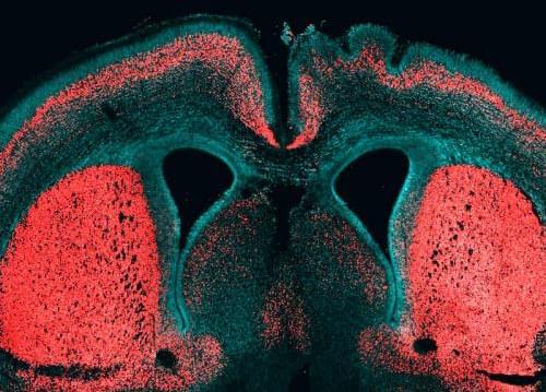
During human evolution, the size of the brain increased, especially in a particular part called the neocortex. The neocortex enables us to speak, dream and think. In search of the causes underlying neocortex expansion, researchers at the Max Planck Institute of Molecular Cell Biology and Genetics in Dresden, together with colleagues at the University Hospital Carl Gustav Carus Dresden, previously identified a number of molecular players. These players typically act cell-intrinsically in the so-called basal progenitors, the stem cells in the developing neocortex with a pivotal role in its expansion.