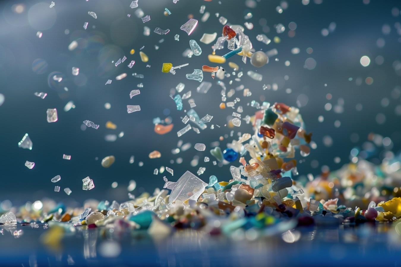
A supplement comprised of dead bacteria seems to remove microplastics, and prevent their absorption into cells, by attracting them to the bacteria’s rough surfaces

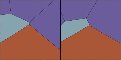
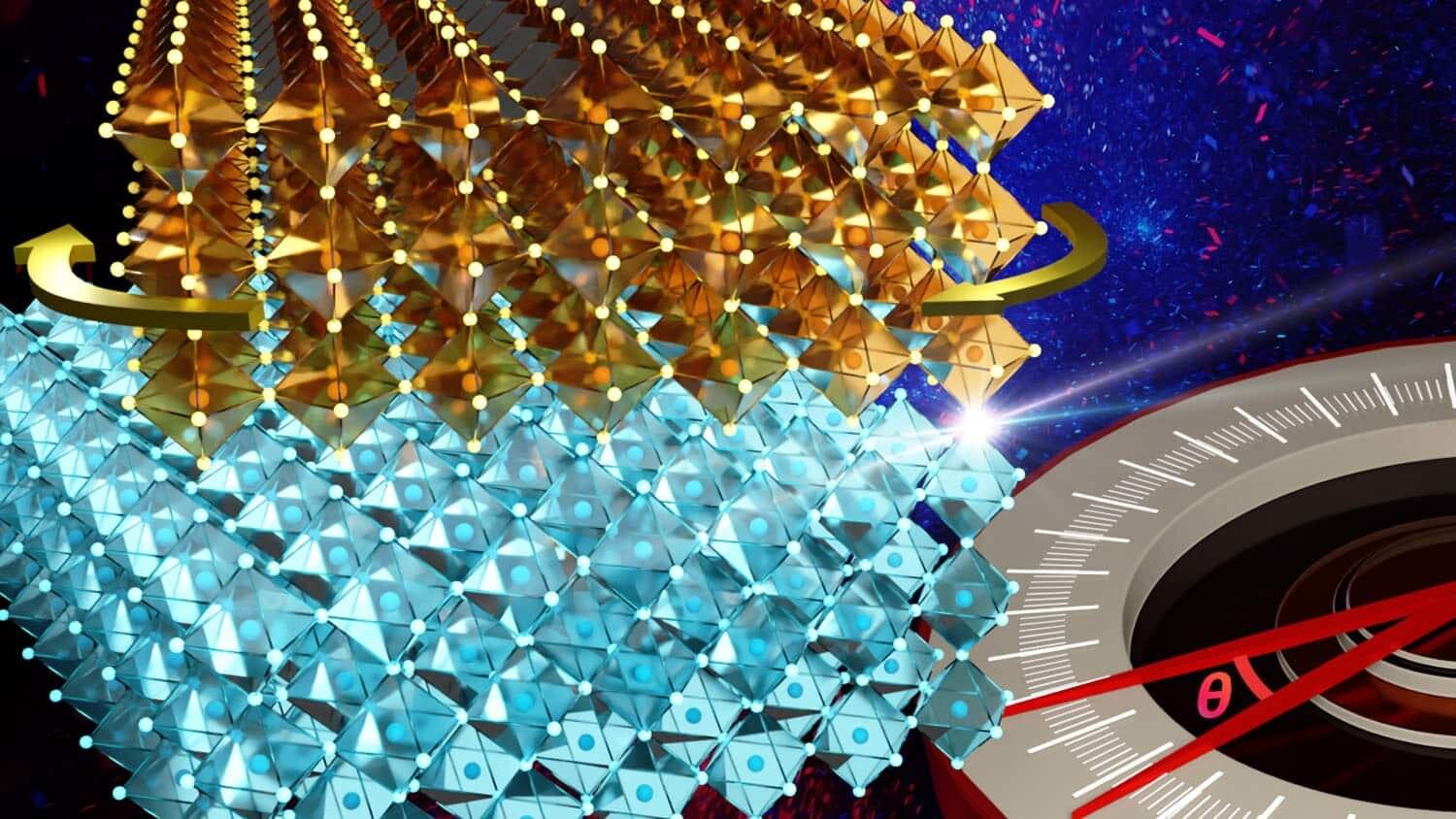
Researchers have shown it is possible to expand the field of twistronics—literally. They have demonstrated a technique that allows them to fabricate oxide twistronic materials at much larger scales while also controlling the twist angles between materials that dictate their structural and electronic properties.
The field of twistronics examines how the angle between layers of two-dimensional (2D) materials affects their electronic properties. The paper, “Deterministic Fabrication of Large-Area, High-Crystallinity Oxide Moiré Superlattices,” is published in the journal ACS Nano.