Proteins are structured like folded chains. These chains are composed of small units of 20 possible amino acids, each labeled by a letter of the alphabet. A protein chain can be represented as a string of these alphabetic letters, very much like a string of music notes in alphabetical notation.
Protein chains can also fold into wavy and curved patterns with ups, downs, turns, and loops. Likewise, music consists of sound waves of higher and lower pitches, with changing tempos and repeating motifs.
Protein-to-music algorithms can thus map the structural and physiochemical features of a string of amino acids onto the musical features of a string of notes.
Without a new legal framework, they could destabilize societal norms.
Autonomous weapon systems – commonly known as killer robots – may have killed human beings for the first time ever last year, according to a recent United Nations Security Council report on the Libyan civil war. History could well identify this as the starting point of the next major arms race, one that has the potential to be humanity’s final one.
Autonomous weapon systems are robots with lethal weapons that can operate independently, selecting and attacking targets without a human weighing in on those decisions. Militaries around the world are investing heavily in autonomous weapons research and development. The U.S. alone budgeted US$18 billion for autonomous weapons between 2016 and 2020.
Indeed, as Gizmodo’s report highlights, many experts believe that these researchers have not actually found evidence of dinosaur DNA. They told the news outlet, for example, that—even under the best circumstances—DNA couldn’t last more than three million years. Let alone more than 100 million. And that the chemicals may have been staining inorganic matter that only looks cellular in nature.
As of now, the most ancient DNA that scientists have been able to sequence was that of a million-year-old woolly mammoth. And the youngest dinosaurs are at least 65 million years old. But if future experiments do confirm this evidence as real, then that really changes things. At least in our fantasies, where reanimated dinosaurs and Ian Malcolms abound.
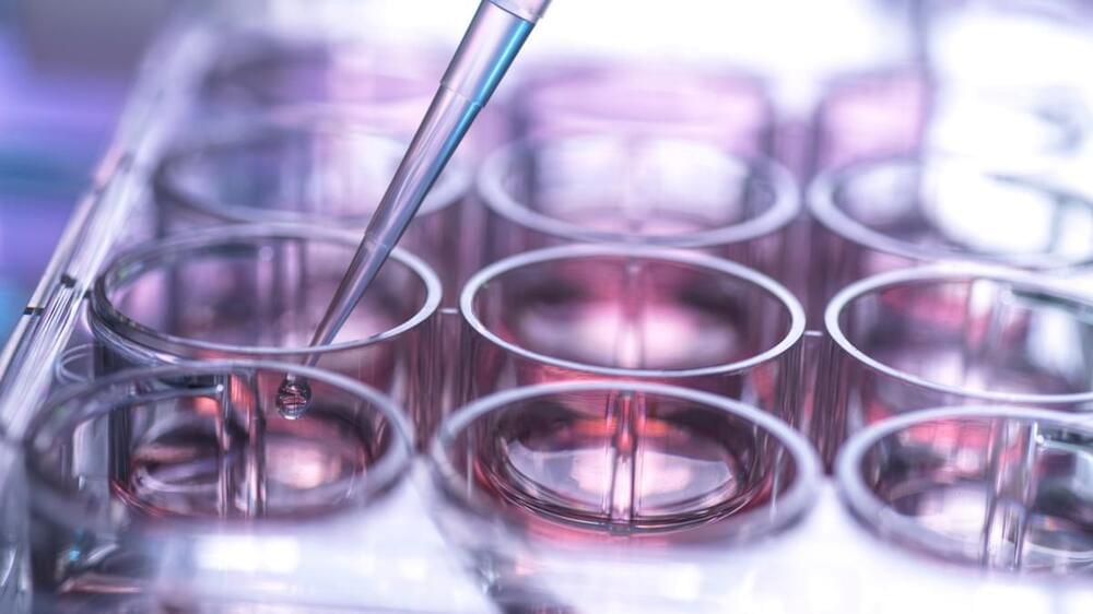
When Dr. Robert Murphy first started researching biochemistry anddrug development in the late 1970s, creating a pharmaceutical compound that was effective and safe to market followed a strict experimental pipeline that was beginning to be enhanced by large-scale data collection and analysis on a computer.
Now head of the Murphy Lab for computational biology at Carnegie Mellon University (CMU), Murphy has watched over the years as data collection and artificial intelligence have revolutionized this process, making the drug creation pipeline faster, more efficient, and more effective.
Recently, that’s been thanks to theapplication of machine learning—computer systems that learn and adapt by using algorithms and statistical models to analyze patterns in datasets—to the drug development process. This has been notably key to reducing the presence of side effects, Murphy says.
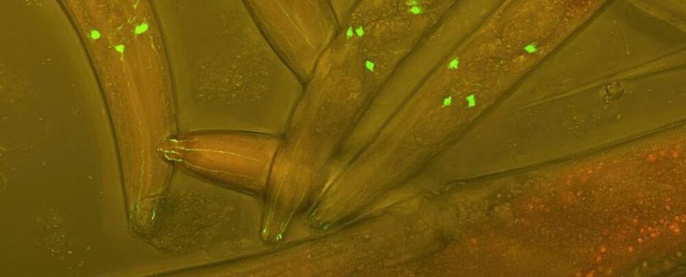
Brains aren’t the easiest of organs to study, what with their delicate wiring and subtle whispering of neurotransmitter messages. Now, this research could be made a little easier, as we’ve learned we can swap some critical chemical systems with the host animal being none the wiser.
In a proof-of-concept study run by a team of US researchers, the microscopic worm Caenorhabditis elegans was genetically gifted pieces of a nervous system taken from a radically different creature – a curious freshwater organism known as Hydra.
The swap wasn’t unlike teaching a specific brain circuit a foreign language, and finding it performs its job just as well as before.
Developing drugs for a range of tauopathies — dr leticia toledo-sherman, senior director, drug discovery, tau consortium, rainwater charitable foundation.
Dr. Toledo-Sherman leads drug discovery activities for an international network of scientists working to develop therapies for Tauopathies, a group of neurodegenerative disorders characterized by the deposition of abnormal Tau protein in the brain.
Previously, Dr. Toledo-Sherman was Director of Medicinal Chemistry and Computer-Aided Drug Design at the CHDI Foundation, leading drug discovery programs for therapeutic development in Huntington’s Disease (HD). At CHDI, she also led a structural biology initiative critical to the understanding of the relationship between structure and biological function of huntingtin, the protein that when mutated causes HD.
Prior to joining CHDI, Dr. Toledo-Sherman was Executive Director of Chemistry at Lymphosign (now part of Pharmascience Inc), a privately held biotechnology company that applied rational design principles to the development of therapeutics for blood cancers. From 2000 to 2,004 she led a multi-site, multidisciplinary team using chemical proteomics and bioinformatics to discover therapeutic targets and to investigate the mechanism of action of drugs.
Autonomous weapon systems – commonly known as killer robots – may have killed human beings for the first time ever last year, according to a recent United Nations Security Council report on the Libyan civil war. History could well identify this as the starting point of the next major arms race, one that has the potential to be humanity’s final one.
Autonomous weapon systems are robots with lethal weapons that can operate independently, selecting and attacking targets without a human weighing in on those decisions. Militaries around the world are investing heavily in autonomous weapons research and development. The U.S. alone budgeted US$18 billion for autonomous weapons between 2016 and 2020.
A possible explanation for life from nonliving material.
Exactly how life first emerged from non-living matter is one of the most enduring mysteries of science. In a new study, Japanese scientists have created self-replicating protocells in the lab, which they say could represent the “missing link” between chemistry and biology.
Primitive Earth was covered with a sludgy mix of chemicals, containing organic molecules that formed the precursors for vital biological components like proteins and amino acids. There are several different hypotheses for how and where life sprang out of this soup, but one of the first ideas was known as chemical evolution, which is what the new study investigated.
“Chemical evolution was first proposed in the 1920s as the idea that life first originated with the formation of macromolecules from simple small molecules, and those macromolecules formed molecular assemblies that could proliferate,” says Muneyuki Matsuo, first author of the study. “However, the origin of molecular assemblies that proliferate from small molecules has remained a mystery for about a hundred years since the advent of the chemical evolution scenario. It has been the missing link between chemistry and biology in the origin of life.”
When people think about the search for life beyond Earth they often think about looking beyond our solar system and even beyond our galaxy. But what about looking closer to home? Titan, Saturn’s largest moon has a dense atmosphere, an internal liquid water ocean, and stable bodies of liquid methane on its surface. While we have not found any evidence of life on Titan, its chemistry and environment make it an interesting place to explore. Europa is a moon of Jupiter with a water-ice crust and liquid ocean underneath. Its atmosphere is very thin, but it’s composed mostly of oxygen.
Zibi (Elizabeth) Turtle is the principal investigator of the Dragonfly mission which will land a drone-like vehicle on Titan to conduct sorties to sample and examine sites around Titan. Morgan Cable is also on the Dragonfly Team, and both Zibi and Morgan are working on the Europa Clipper mission which will perform reconnaissance of Europa to investigate whether it could have conditions suitable for life. Join Zibi and Morgan, along with SETI Institute planetary astronomer Franck Marchis for their discussion about what makes Titan and Europa such intriguing places to search for clues about the origins of life in our solar system.
If you like science, support the SETI Institute! We’re a non-profit research institution whose focus is understanding the nature and origins of life in the universe. Donate here: https://seti.org/donate.
Learn more about the SETI Institute and stay up-to-date on awesome science:
An international team of researchers has used liquid gallium to create an antiviral and antimicrobial coating and tested it on a range of fabrics, including facemasks. The coating adhered more strongly to fabric than some conventional metal coatings, and eradicated 99% of several common pathogens within five minutes.
“Microbes can survive on the fabrics hospitals use for bedding, clothing and face masks for a long time,” says Michael Dickey, co-corresponding author of a paper on the work and Camille & Henry Dreyfus Professor of Chemical and Biomolecular Engineering at North Carolina State University. “Metallic surface coatings such as copper or silver are an effective way to eradicate these pathogens, but many metal particle coating technologies have issues such as non-uniformity, processing complexity, or poor adhesion.”
Dickey and colleagues from NC State, Sungkyunkwan University (SKKU) in Korea and RMIT University in Australia set out to develop a simple, cost-effective way to deposit metal coatings on fabric.