Toggle light / dark theme

Machine learning enables real-time analysis of iron oxide thin film growth in reactive magnetron sputtering

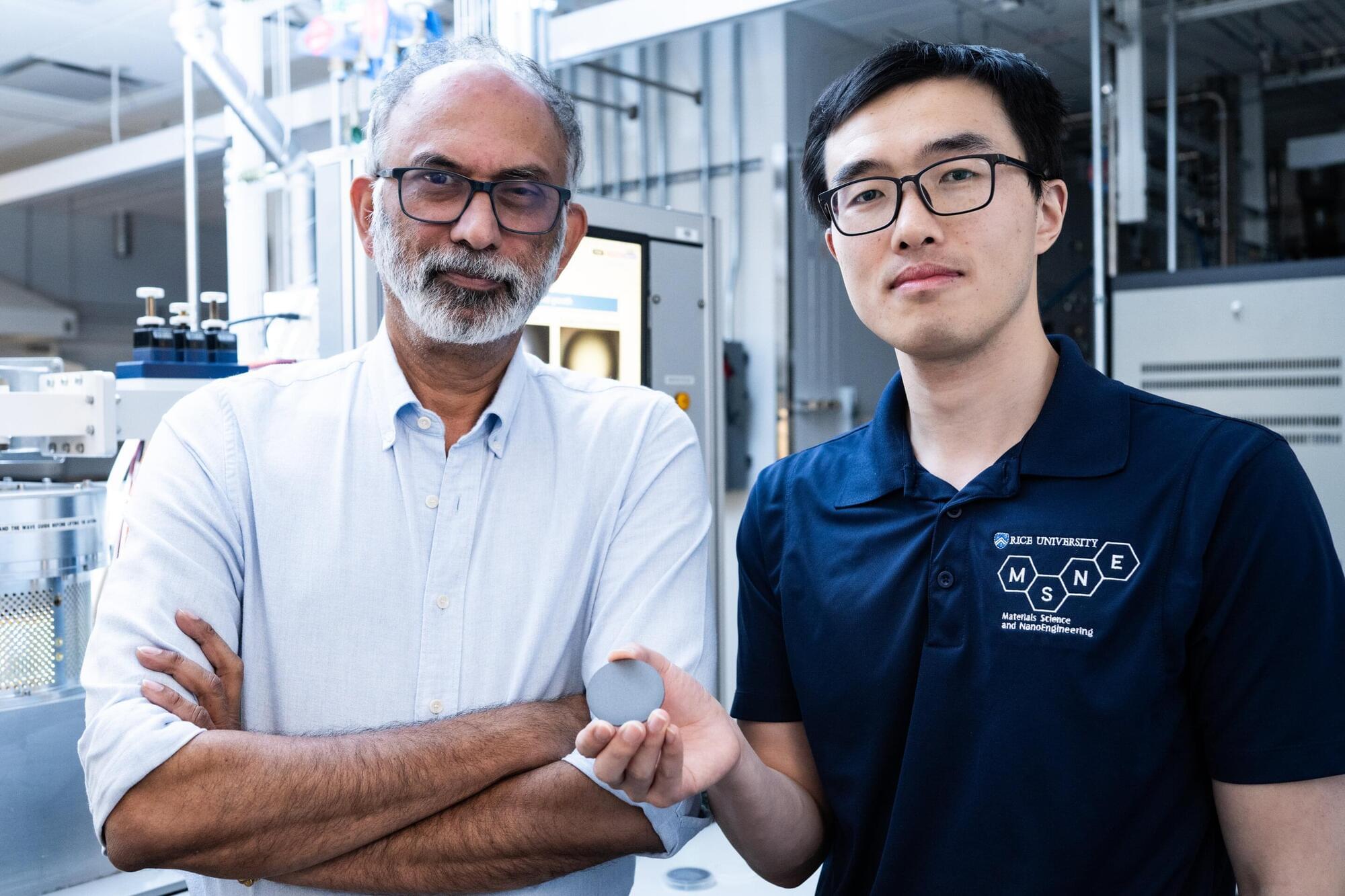
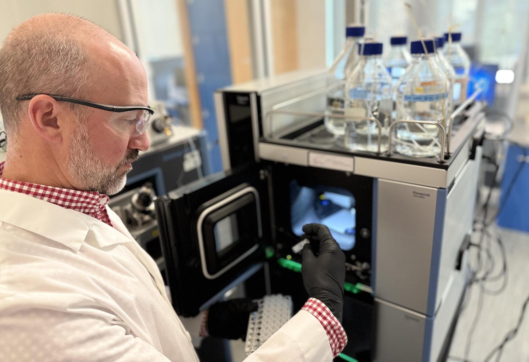
Researchers at University of Tsukuba have developed a technology for real-time estimation of the valence state and growth rate of iron oxide thin films during their formation. This novel technology was realized by analyzing the full-wavelength data of plasma emission spectra generated during reactive sputtering using machine learning. It is expected to enable high-precision control of the film deposition process.

Metal oxide and nitride thin films are commonly used in and energy materials. Reactive sputtering is a versatile technique for depositing thin films by reacting a target metal with gases such as oxygen or nitrogen. A challenge with this process is the transitioning of the target surface between metallic and compound states, causing large fluctuations in film growth rate and composition. At present, there are limited effective methods for real-time monitoring of a material’s chemical state and deposition rate during film formation.

A machine learning technique based on was employed to examine massive emission spectra generated within a reactive sputter plasma. This analysis focused on assessing the state of thin film formation. The results, published in Science and Technology of Advanced Materials: Methods, indicated that the valence state of iron oxide was accurately identified using only the first and second principal components of the spectra. In addition, the film growth rate was predicted with high precision.

RNA modifications control how stem cells develop into retinal cells, research demonstrates

Cells contain a blueprint in the form of DNA that dictates what they can make. This blueprint is converted into a message (mRNA), which is then converted into a protein. Although DNA remains the same in all cells, how it is read depends on specific signals that can change the DNA itself, mRNA or proteins. These signals are often in the form of chemical modifications.

Crystal-free mechanoluminescence illuminates new possibilities for next-generation materials

In the 17th century, Francis Bacon described a simple experiment—scraping and fracturing hard sugar in the dark to see sparks of light. This phenomenon is called mechanoluminescence (ML) or triboluminescence (TL), the process of materials emitting light under mechanical stimulation, like grinding or crushing. Usually, ML properties of luminescent compounds are observed in rigid crystalline systems, which limits their real-world applications.

Now, researchers at the Okinawa Institute of Science and Technology (OIST) have found a way to generate ML in non-crystalline materials, bringing a new wave of potential applications in engineering, industrial safety and beyond.

“Mechanical stimulation of crystals causes fractures. As the crystals are damaged and break down in size, they also start to lose their ML properties, which vastly restricts their application. In , ML is highly dependent on structure and packing, adding complex design requirements. That’s why we were interested in amorphous ML materials with longer-lasting luminescence,” explains Professor Julia Khusnutdinova, head of the Coordination Chemistry and Catalysis Unit at OIST.

Electrons can now be controlled to build smarter quantum devices

Auburn University scientists have developed a new class of materials that lets researchers precisely control free electrons, a breakthrough that could reshape the future of computing and chemical manufacturing.

Their study introduces a material system that allows fine-tuned control over how electrons behave within matter, potentially paving the way for faster computers, smarter machines, and more efficient industrial processes.

Chemists discover antibiotic for drug-resistant bacteria ‘hiding in plain sight’

Chemists from the University of Warwick and Monash University have discovered a promising new antibiotic that shows activity against drug-resistant bacterial pathogens, including MRSA and VRE

Antimicrobial resistance (AMR) is one of the world’s most urgent health challenges, with the WHO’s new report showing there are ‘too few antibacterials in the pipeline. Most of the ‘low-hanging fruit’ has already been found, and the limited commercial incentives deter investment in antibiotic discovery.

In a study published in the Journal of the American Chemical Society, researchers from the Monash Warwick Alliance Combating Emerging Superbug Threats Initiative have discovered a promising new antibiotic—pre-methylenomycin C lactone.

Imaging technique maps fleeting intermediates in hydrogen electrocatalysis

Electrocatalytic transformations not only require electrical energy—they also need a reliable middleman to spark the desired chemical reaction. Surface metal-hydrogen intermediates can effectively produce value-added chemicals and energy conversion, but, given their low concentration and fleeting lifespan, they are difficult to characterize or study in depth, especially at the nanoscale.

All-solid-state battery researchers reveal key insights into degradation mechanisms

Researchers from UNIST, Seoul National University (SNU), and POSTECH have made a significant breakthrough in understanding the degradation mechanisms of all-solid-state batteries (ASSBs), a promising technology for next-generation electric vehicles and large-scale energy storage.

Jointly led by Professor Donghyuk Kim at UNIST’s School of Energy and Chemical Engineering, Professor Sung-Kyun Jung at SNU’s School of Transdisciplinary Innovations, and Professor Jihyun Hong from POSTECH, their study reveals that interfacial chemical reactions play a critical role in structural damage and performance decline in sulfide-based ASSBs. The findings are published in Nature Communications.

Unlike that rely on flammable liquid electrolytes, ASSBs use non-flammable solid electrolytes, offering enhanced safety and higher energy density. However, challenges such as interface instability and microstructural deterioration have impeded their commercialization. Until now, the detailed understanding of how these phenomena occur has remained limited.

Mesoscale volumetric fluorescence imaging at nanoscale resolution by photochemical sectioning

I first explored this amazing work back when it was a preprint! Wang et al. herein developed VIPS (volumetric imaging via photochemical sectioning), a way of using UV light to remove layers of expanded tissue-hydrogel, allowing combination of high-resolution lattice-light sheet microscopy with expansion microscopy. Link: [ https://www.science.org/doi/10.1126/science.adr9109]

In my opinion, this technology has enormous future promise for high-throughput connectomics! They will need to improve their labeling density so that higher expansion factors can be used, but this problem is well-studied and I think the issue will likely be solvable with additional resources/effort.


Optical nanoscopy of intact biological specimens has been transformed by recent advancements in hydrogel-based tissue clearing and expansion, enabling the imaging of cellular and subcellular structures with molecular contrast. However, existing high-resolution fluorescence microscopes are physically limited by objective-to-specimen distance, which prevents the study of whole-mount specimens without physical sectioning. To address this challenge, we developed a photochemical strategy for spatially precise sectioning of specimens. By combining serial photochemical sectioning with lattice light-sheet imaging and petabyte-scale computation, we imaged and reconstructed axons and myelin sheaths across entire mouse olfactory bulbs at nanoscale resolution.

/* */