MIT neuroscientists have made a breakthrough in treating fragile X syndrome by leveraging a novel neurotransmitter signaling pathway. By targeting a specific subunit of NMDA receptors, they successfully reduced excessive protein synthesis in the brain, a hallmark of the disorder. Their approach, tested in fragile X model mice, not only corrected molecular imbalances but also improved synaptic function and reduced disease symptoms.
Category: biotech/medical – Page 873
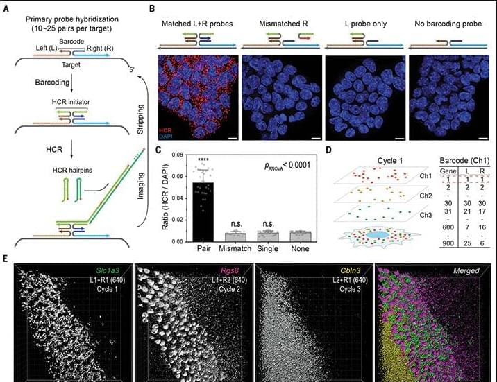
Deep-tissue transcriptomics and subcellular imaging at high spatial resolution
Limited color channels in fluorescence microscopy have long constrained spatial analysis in biological specimens. Here, we introduce cycle Hybridization Chain Reaction (HCR), a method that integrates multicycle DNA barcoding with HCR to overcome this limitation. cycleHCR enables highly multiplexed imaging of RNA and proteins using a unified barcode system. Whole-embryo transcriptomics imaging achieved precise three-dimensional gene expression and cell fate mapping across a specimen depth of ~310 μm. When combined with expansion microscopy, cycleHCR revealed an intricate network of 10 subcellular structures in mouse embryonic fibroblasts. In mouse hippocampal slices, multiplex RNA and protein imaging uncovered complex gene expression gradients and cell-type-specific nuclear structural variations.
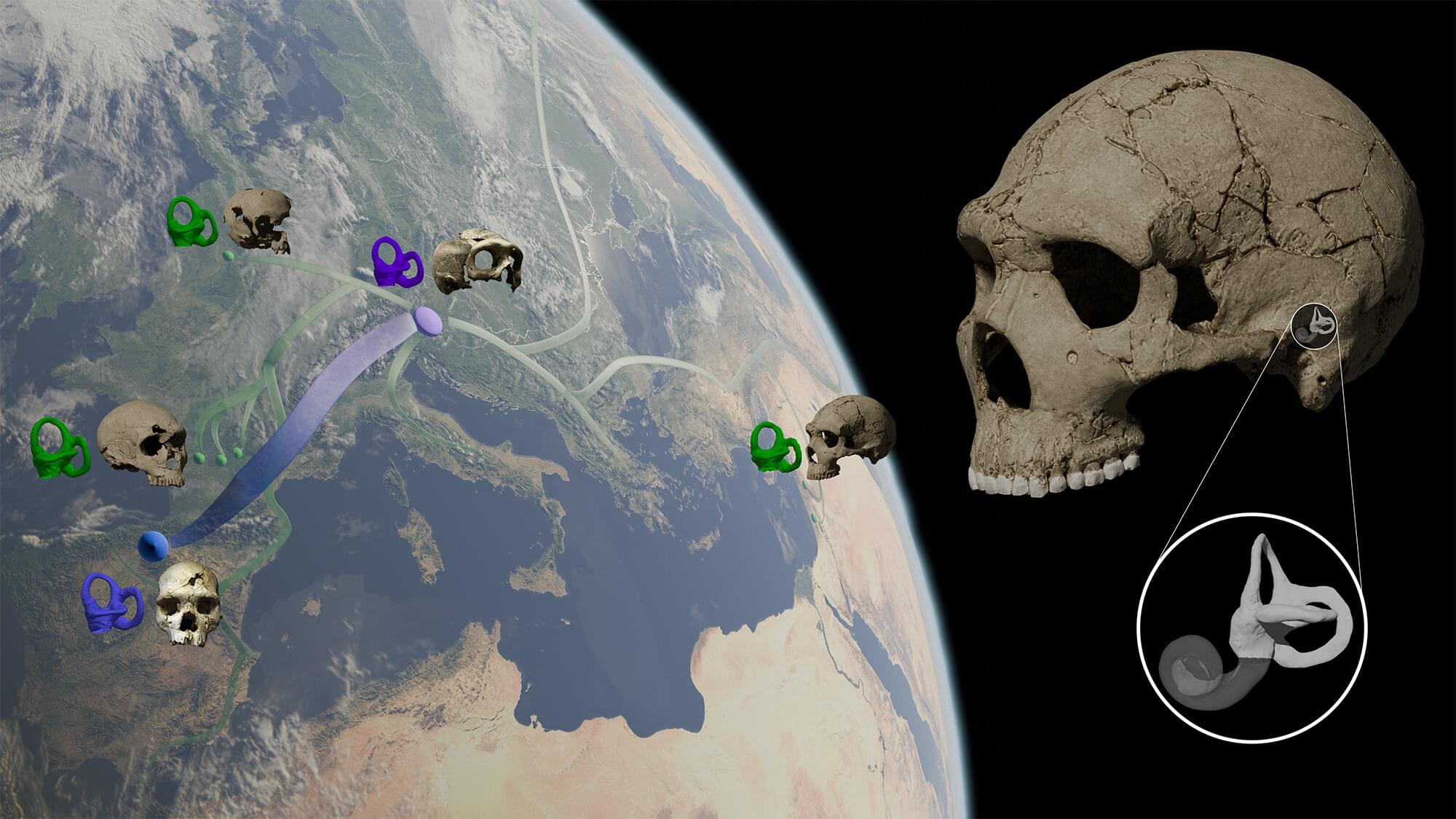
The inner ear of Neanderthals reveals clues about their enigmatic origin
New research on the inner ear morphology of Neanderthals and their ancestors challenges the widely accepted theory that Neanderthals originated after an evolutionary event that implied the loss of part of their genetic diversity. The findings, based on fossil samples from Atapuerca (Spain) and Krapina (Croatia), as well as from various European and Western Asian sites have been published in Nature Communications.
Neanderthals emerged about 250,000 years ago from European populations—referred to as “pre-Neanderthals”—that inhabited the Eurasian continent between 500,000 and 250,000 years ago. It was long believed that no significant changes occurred throughout the evolution of Neanderthals, yet recent paleogenetic research based on DNA samples extracted from fossils revealed the existence of a drastic genetic diversity loss event between early Neanderthals (or ancient Neanderthals) and later ones (also referred to as “classic” Neanderthals).
Technically known as a “bottleneck,” this genetic loss is frequently the consequence of a reduction in the number of individuals in a population. Paleogenetic data indicate that the decline in genetic variation took place approximately 110,000 years ago.
The World’s First Faceless, Synthetic ‘Human’ Is Straight-Up Nightmare Fuel
We knew this day would come. For years now, scientists have been trying to make Westworld a reality with their advances in android robots.
From lab-grown muscle tissue to robots with bones, ligaments and tendons, android robots have rapidly been getting more and more lifelike.
Now, a company named Clone Robotics has created something they are calling the Protoclone – a “faceless, anatomically accurate, synthetic human with over 200 degrees of freedom, over 1,000 Myofibers, and over 200 sensors.” Sound terrifying? Just wait until you see video of it twitching and spasming its way into action.

Stopping cancer cells from infiltrating nervous system
About 15 years ago, Stanford Medicine neuro-oncologist Michelle Monje, MD, PhD, began to suspect that the brain tumors she studied were doing something strange. Cancer cells sometimes copycat their healthy counterparts, so Monje and her team weren’t surprised to uncover simple parallels between healthy and malignant brain cells. The cancer’s biological “borrowing” was similar to a symphony-goer who whistles the theme from a concerto on the bus ride home.
But the team’s data hinted that these brain tumors were orchestrating something much more complex. Instead of just humming the themes of healthy brain biology, the research suggested the tumors could round up many important cell-signaling instruments — the microscopic equivalents of, say, violins, cellos, flutes and trombones — and use them to play a score of its own.
In physiologic terms, Monje’s team gradually demonstrated, certain cancer cells form working electrical connections with nearby nerves. The tumors wire themselves neatly into the brain’s electrical apparatus, then use healthy nerves’ signals for their own purposes — to drive malignant growth. These cancers also hijack the machinery of learning to strengthen connections with the healthy brain and further enhance their ability to multiply.
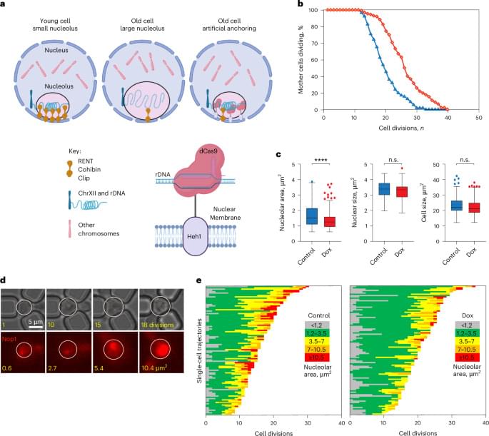
A mortality timer based on nucleolar size triggers nucleolar integrity loss and catastrophic genomic instability
Gutierrez and Tyler investigate the limits of replicative lifespan in yeast. The authors show that nucleolar expansion during aging is a mortality timer. Enlargement of nucleoli beyond a defined size alters their biophysical properties; normally excluded DNA repair protein enter, causing aberrant rDNA recombination, genome instability and death.
Scientists create ‘gene switch’ to power cell therapy for diabetes
Researchers have developed a new, highly effective “gene switch” to deliver targeted cell therapy.
The ETH Zurich team states that this cell therapy has the potential to offer a more precise and personalized treatment for diabetes.
Diabetes is a major global health concern, classified as a metabolic disease and affecting about one in ten individuals.

Incyte, Genesis Therapeutics Partner on AI-Based Small Molecule Collaboration
Incyte will partner with Genesis Therapeutics to research, discover, and develop small molecule treatments through a collaboration that could generate at least up to $620 million for Genesis, an artificial intelligence (AI)-based drug developer.
The companies have agreed to discover and optimize at least two initial small molecule programs through Genesis’s AI platform, Genesis Exploration of Molecular Space (GEMS). GEMS is designed to generate and optimize molecules for complex targets by integrating proprietary AI methods that include language models, diffusion models, and physical machine learning (ML) simulations.
Incyte has been granted exclusive rights for potential clinical development and commercialization of the products to be developed through the collaboration.

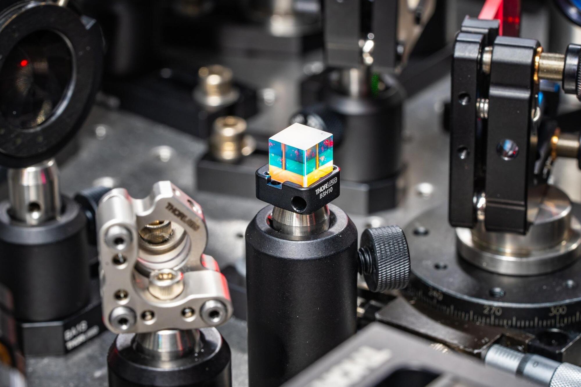
Can You Hear a Virus? Scientists Use Light To Listen to the Sounds of Life
Elad Harel is used to shining a light on the mysteries of the natural world.
Working at the cutting edge of ultrafast spectroscopy—the use of short laser pulses to analyze molecular dynamics—the Michigan State University associate professor seeks to uncover how microscopic phenomena impact large complex systems.
One promising frontier Harel has been working on is the development of new methods of microscopy that will allow researchers to observe molecular and atomic landscapes in motion rather than through static imagery. Such work has earned Harel MSU’s 2023 Innovation of the Year award, as well as MSU’s first-ever grant from the W.M. Keck Foundation.