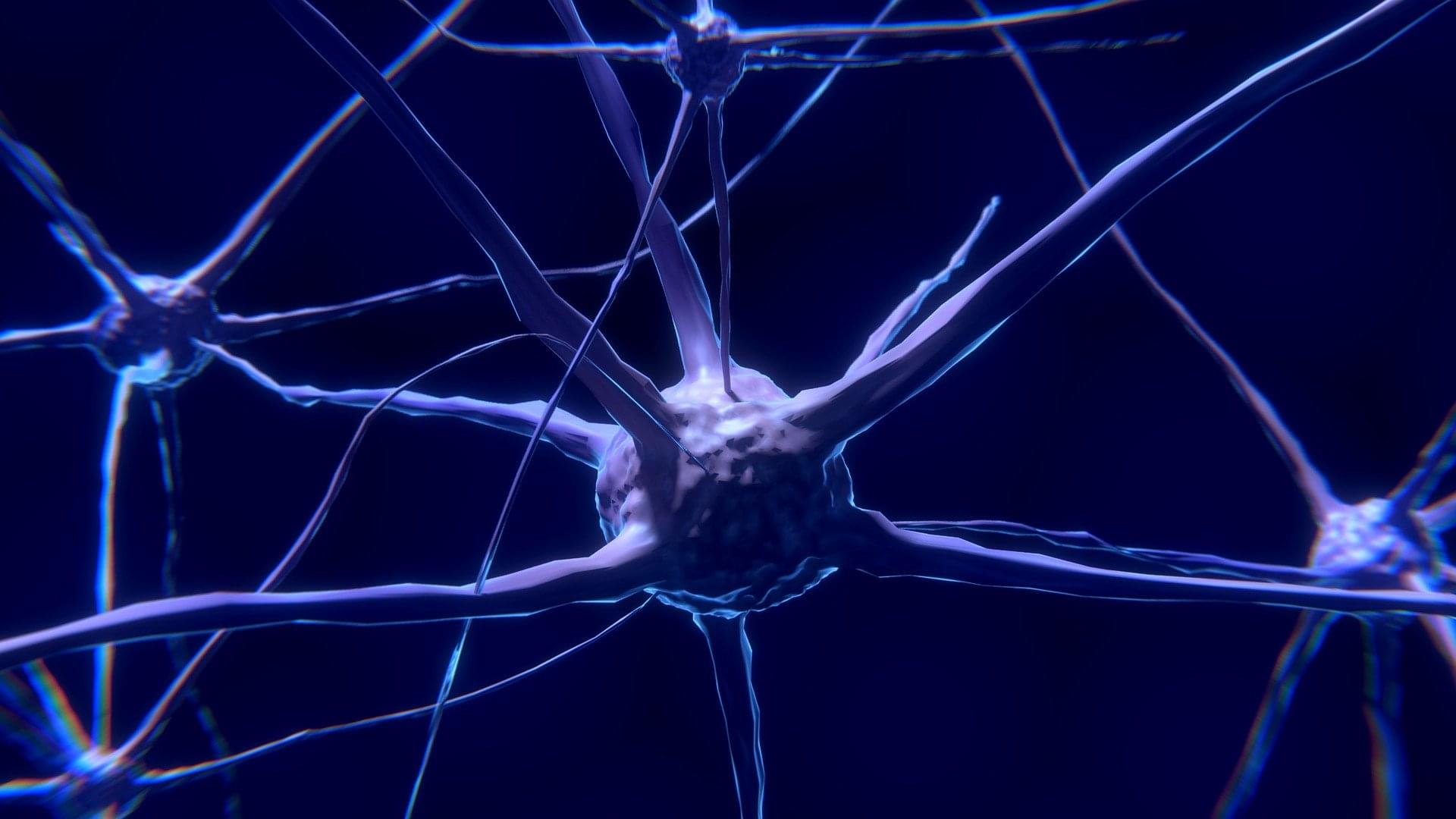
Citrate is essential for the metabolism and development of neurons. A membrane transport protein called SLC13A5 plays a central role in this process and has previously been linked to a particularly severe form of epileptic encephalopathy.
Building on data from the recently completed RESOLUTE and REsolution flagship projects, scientists at CeMM have comprehensively studied the function and structure of the membrane transporter SLC13A5, experimentally investigating 38 mutant variants.
Their findings, published in Science Advances, shed new light on the mechanisms of this disease and lay the foundation for further research into epilepsy and other disorders.
Our brain makes decisions based on direct associations between stimuli in our environment, but it often also does so based on events that initially appear unrelated. How does it achieve this? A recent study by the Cellular Mechanisms in Physiological and Pathological Behavior Research Group at the Hospital del Mar Research Institute, published in Proceedings of the National Academy of Sciences, offers new insights into this process and identifies the brain areas involved.
Using observations in mice, led primarily by first author and Ph.D. student José Antonio González Parra and supervised by Dr. Arnau Busquets, the research team was able to determine the mechanisms involved in how the brain makes decisions based on indirect associations between different stimuli. That is, instead of directly associating a specific stimulus with a rewarding or aversive situation, the brain establishes connections between two or more stimuli.
Dr. Busquets explains, “The project aims to understand how the brain enables us to make decisions based on indirect relationships between stimuli in our environment.”
Over the past decades, some lawyers have started using brain imaging scans as evidence during criminal trials, to provide a possible explanation for the criminal behavior of defendants. This was justified by recent neuroscientific studies, which found that some people who commit crimes present differences in specific parts of the brain. Yet a key question remains: are these brain changes causal, compensatory or incidental to the behavior?
To answer this question, researchers at Brigham and Women’s Hospital, Harvard Medical School and other institutes in the U.S. analyzed the locations of brain injury temporally associated with a new onset of criminality.
They found evidence suggesting that lesions to a specific white matter tract could be causally implicated in the behavior of individuals who start committing crimes after injury.
This retrospective cohort study was conducted using data from TriNetX, a multi-institutional health research network. Using the TriNetX platform, we accessed deidentified electronic health records from over 212 million patients across 120 major health care organizations.9 The built-in analytic functions of TriNetX enable patient-level analyses while ensuring that only population-level data are reported.
This study was approved by WCG Clinical, which granted a waiver to TriNetX as a federated network and was deemed exempt from informed consent owing to the use of existing, non–human participant data that were deidentified per the US Health Insurance Portability and Accountability Act privacy rule. The study followed the Strengthening the Reporting of Observational Studies in Epidemiology (STROBE) reporting guideline.
We included patients with cirrhosis (International Statistical Classification of Diseases and Related Health Problems, Tenth Revision [ICD-10] codes K74.6 and K74.69), who were taking furosemide (RxNorm [National Library of Medicine] code 4603) and spironolactone (RxNorm code 9997) between January 2013 and July 2021. For patients receiving an SGLT-2 inhibitor (Anatomical Therapeutic Chemical code A10BK), the index event was defined as the date on which they were concurrently prescribed spironolactone, furosemide, and an SGLT-2 inhibitor. For the control group, the index event was the date on which they were prescribed concurrent spironolactone and furosemide but not an SGLT-2 inhibitor. Each patient was followed up for 3 years from the index event, with follow-up ending on July 11, 2024.
Why do some patients with precursors to bone marrow cancer never develop the disease? Researchers from the Department of Forensic Medicine at Aarhus University have discovered that some cells enter a dormant state and create a defense against cancer—a breakthrough that could lead to early treatment.
In a new study published in the journal Leukemia, researchers investigated multiple myeloma, a serious bone marrow cancer that arises in plasma cells.
Before the disease emerges, patients always have a precursor condition, either MGUS (monoclonal gammopathy of undetermined significance) or SMM (smoldering multiple myeloma). These conditions are not cancer themselves but are associated with an increased risk of developing bone marrow cancer—approximately 1% and 10% increased risk per year, respectively.
What if a blood test could reveal the pace of our aging—and the diseases that may lie ahead? The labs of Profs. Liran Shlush and Amos Tanay at the Weizmann Institute of Science have been conducting in-depth studies into the biology of blood to better understand the aging process and why some people become more susceptible to disease over the years.
Their research teams, made up of physicians, biologists and data scientists, have been tracking changes in the blood-forming stem cells, including the emergence of genetic changes in these cells in about one-third of people over the age of 40. These changes not only increase the risk of blood cancers such as leukemia, but have also been linked to heart disease, diabetes and other age-related conditions.
In a new study published today in Nature Medicine, Shlush and Tanay present findings that may lead to an innovative blood test for detecting a person’s risk of developing leukemia. This test may potentially replace the invasive diagnostic procedure of bone marrow sampling.
Gene therapy—once something out of science fiction—is now being used in real hospitals to treat real people. Gene editing has become a conversation of not only treating rare diseases but also about access, fairness, and how much control we should have over our biology.
Genes are sections of DNA that act like instruction manuals telling our cells how to build proteins. Proteins perform vital function like energy use, cellular communications, immunity and cell repair. So when people say “We are what our genes make us,” it’s because these gene-coded proteins guide our growth, health, and behaviour.
Sometimes, typos appear in these instruction manuals. They are called genetic mutations. While many mutations are harmless, some affect the protein made from the mutated gene and disrupt how the cell functions. Some cause serious diseases like cystic fibrosis, muscular atrophy or certain cancers.
Join us for an exclusive 1-hour conversation with Dr. Eriona Hysolli, the visionary scientist bridging de-extinction technology and the future of human reproduction. Recognized by Time100 Next for her groundbreaking work reviving the woolly mammoth, Dr. Hysolli brings a unique perspective to reproductive biotechnology that you won’t find anywhere else.
In this informal Q&A session, we’ll explore how cutting-edge technologies originally developed for species conservation are now revolutionizing human fertility treatments. Dr. Hysolli will share insights on: The latest breakthroughs in synthetic embryos and artificial wombs. How in vitro gametogenesis could transform infertility treatment. Lessons from mammoth de-extinction that apply to human reproductive health. The intersection of genome engineering and fertility solutions. Near-term commercial applications in reproductive biotechnology.
Drawing from her pioneering work at Yale, George Church’s lab at Harvard, and as Head of Biological Sciences at Colossal Biosciences, Dr. Hysolli offers a rare glimpse into technologies that could redefine human reproduction within the next decade.
The session will feature a moderated discussion followed by audience Q&A. Whether you’re an investor, entrepreneur, healthcare professional, or simply fascinated by the future of fertility, this conversation will provide essential insights into one of biotechnology’s most promising frontiers.
Scientists have created tiny disk-shaped particles that can swim on their own when hit with light, akin to microscopic robots that move through a special liquid without any external motors or propellers.
Published in Advanced Functional Materials, the work shows how these artificial swimmers could one day be used to deliver cargo in a variety of fluidic situations, with potential applications in drug delivery, water pollutant clean-up, or the creation of new types of smart materials that change their properties on command.
“The essential new principles we discovered—how to make microscopic objects swim on command using simple materials that undergo phase transitions when exposed to controllable energy sources—pave the way for applications that range from design of responsive fluids, controlled drug delivery, and new classes of sensors, to name a few,” explained lead researcher Juan de Pablo.