Aging isn’t just about living longer. It’s about staying healthy while doing it. And a new study just brought us one step closer.

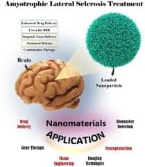
This review explores the transformative potential of nanotechnology in the treatment and diagnosis of amyotrophic lateral sclerosis (ALS), a progressive neurodegenerative disorder characterized by motor neuron degeneration, muscle weakness, and eventual paralysis. Nanotechnology offers innovative solutions across various domains, including targeted drug delivery, neuroprotection, gene therapy and editing, biomarker detection, advanced imaging techniques, and tissue engineering. By enhancing the precision and efficacy of therapeutic interventions, nanotechnology facilitates key advancements such as crossing the blood-brain barrier, targeting specific cell types, achieving sustained therapeutic release, and enabling combination therapies tailored to the complex pathophysiology of ALS.
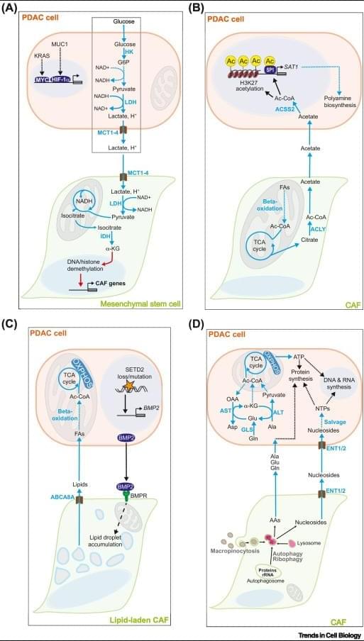
Metabolic crosstalk – the exchange of metabolites between cancer cells and non-malignant cells in the tumor microenvironment (TME) – contributes to the aggressiveness of pancreatic ductal adenocarcinoma (PDAC) through a diverse array of mechanisms.
Under the selection pressure imposed by chemical stressors (acidosis, hypoxia) and scarcity of essential nutrients in the TME, PDAC cells establish mutually fitness-enhancing metabolic crosstalk pathways with cancer-associated fibroblasts, tumor-associated macrophages, and other stromal cells.
PDAC cell metabolism inhibits the activity of cytotoxic T lymphocytes and natural killer cells by outcompeting them for essential nutrients (glucose, amino acids, nucleosides, vitamins) and by flooding the TME with immunosuppressive metabolites (lactate, kynurenine, adenosine, and others).
Critical nodes of tumorigenic metabolic crosstalk pathways (enzymes and cell membrane transporters) are readily druggable and likely non-essential for healthy tissues. https://sciencemission.com/Tumor%E2%80%93stromal-metabolic-crosstalk-in-PC
Pancreatic ductal adenocarcinoma (PDAC) is an aggressive malignancy with a dire prognosis. Standard-of-care chemotherapy regimens offer marginal survival benefit and carry risk of severe toxicity, while immunotherapy approaches have uniformly failed in clinical trials. Extensive desmoplasia in the PDAC tumor microenvironment (TME) disrupts blood flow to and from the tumor, thereby creating a nutrient-depleted, hypoxic, and acidic milieu that suppresses the function of antitumor immune cells and imparts chemotherapy resistance. Additionally, recent seminal studies have demonstrated crucial roles for metabolic crosstalk – the exchange of metabolites between PDAC cells and stromal cell populations in the TME – in establishing and maintaining core malignant behaviors of PDAC: tumor growth, metastasis, immune evasion, and therapy resistance.
We are currently facing the possibility of achieving immortality for humans by 2030. This prediction comes from renowned futurist Ray Kurzweil, who has a history of making accurate predictions. He anticipates that with the ongoing progress in genetics, robotics, and nanotechnology, we will soon have nanobots coursing through our bloodstream, which could enable us to live forever. It’s truly remarkable to consider that this could be a reality within just seven years.
Nanobots, which are small robots sized between 50–100 nm in width, are currently being used in various clinical medical applications. They are used in research as DNA probes, imaging materials for cells, and targeted delivery vehicles for cells. According to Kurzweil, nanobots represent the future of medicine.
They will be capable of repairing our bodies at a cellular level, making us resistant to diseases, aging, and, ultimately death. Additionally, he theorizes that humans may be able to transfer their consciousness into digital form, leading to immortality.
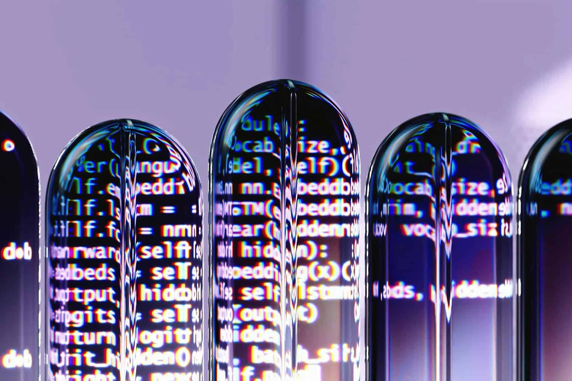
If you thought storing data inside DNA was cool, here’s something even more fascinating. Scientists at the University of Texas at Austin (UT Austin) have invented a way to store digital information inside synthetic polymer molecules. In short, they are transforming tiny bits of plastic into memory banks.
They even used their molecular system to encode a complex 11-character password and then decode it using only electrical signals, without any power, and the expensive and bulky tools typically used for reading molecular data.
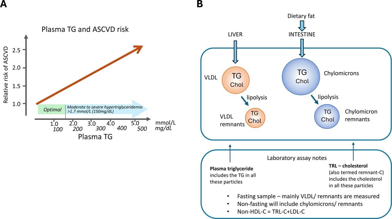
Ischaemic heart disease remains the main cause of death worldwide. 1 Within its multifactorial aetiology low-density lipoprotein (LDL) and other apolipoprotein (apo) B-containing lipoproteins play a central, causal role, promoting the development of the underlying process of atherosclerosis. The use of statins and other drugs—ezetimibe, proprotein convertase subtilisin/kexin type 9 (PCSK9) inhibitors, bempedoic acid—to lower LDL is a central strategy in the prevention of atherosclerotic cardiovascular disease (ASCVD) in both primary and secondary settings. 2 However, in many individuals, a substantial ASCVD risk remains after LDL-cholesterol (LDL-C) goal achievement, and elevated plasma triglyceride (TG) is recognised as an important component of this residual risk. 3 Plasma TG, or more specifically TG-rich lipoproteins (TRL), is therefore an additional target for lipid-lowering therapy. Outcome studies of TG lowering using classical drugs such as fibrates and high-dose niacin when added to statins failed to demonstrate further ASCVD risk reduction, although retrospective analyses suggest that subgroups characterised by high TG and low high-density lipoprotein (HDL) may have positive results. 4–7 An alternative approach, treatment with high-dose eicosapentaenoic acid (EPA), has been shown to reduce cardiovascular risk in patients with (and without) hypertriglyceridaemia who are on statins. 8–10
This review explores the concepts behind, and practical implementation of, an evidence-based therapeutic strategy that tailors further intervention according to the plasma lipid profile in patients on standard statin therapy who are often undertreated. 11
Genetic analyses provide robust evidence that elevated TG is a causal risk factor for ASCVD 12 13 and underpin the finding from epidemiological studies that raised TG levels are positively and linearly related to cardiovascular risk (figure 1A). 14 15 The importance of these observations is that they reveal an often unaddressed major risk factor that is of particular relevance in people with obesity or type 2 diabetes in whom TG levels are frequently elevated. 16 Further, outcome trials have shown that elevated TG levels (again especially in type 2 diabetics) are associated with high residual cardiovascular risk in statin-treated patients with established cardiovascular disease, even if they have well-controlled LDL-C. 17–19.
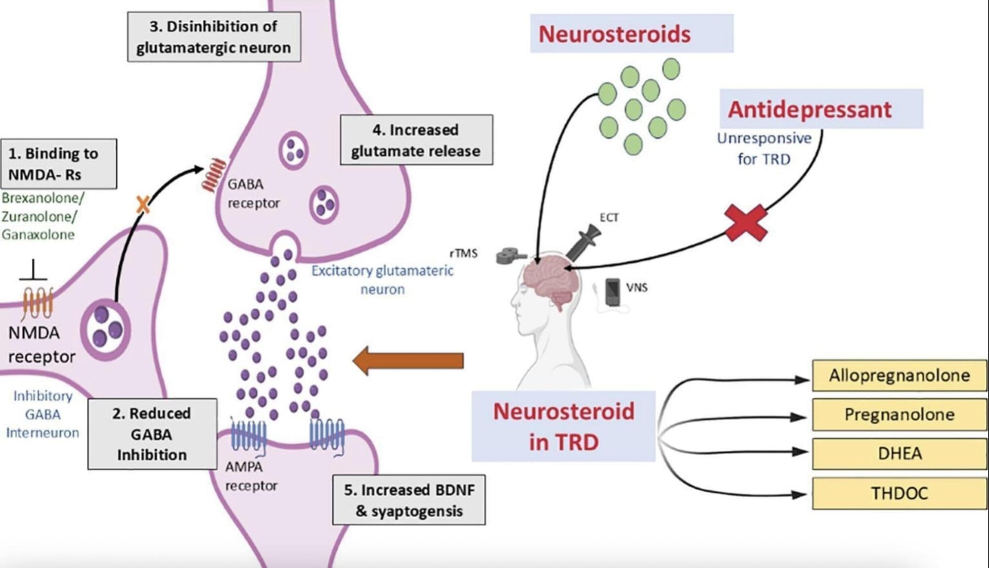
Depression, characterized by persistent sadness, hopelessness and a lack of interest in previously enjoyed activities, is one of the most common mental health disorders. Recent estimates by the World Health Organization (WHO) suggest that approximately 5% of the global population suffers from depression.
For decades, researchers have been trying to devise safe and effective treatments for depression that cause minimal or no side effects. This led to the introduction of a wide range of treatment strategies, ranging from psychotherapy and alternative medicine to a wide range of pharmacological drugs, including selective serotonin reuptake inhibitors (SSRIs), serotonin-norepinephrine reuptake inhibitors (SNRIs), tricyclic antidepressants (TCAs), monoamine oxidase inhibitors (MAOIs) and atypical antidepressants.
Most people diagnosed with depression eventually find a suitable treatment for them via a trial-and-error process, ultimately leading to their recovery. Some individuals, however, can experience severe depression for prolonged periods of time, finding that no treatment ultimately eases their symptoms.
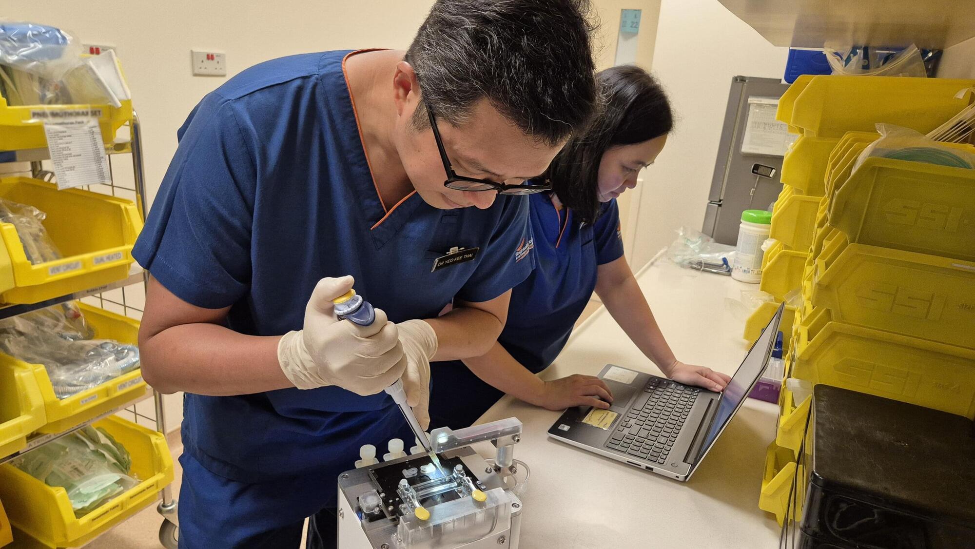
Researchers have developed a first-of-its-kind device to profile the immune function of newborns. Using a single drop of blood, the BiophysicaL Immune Profiling for Infants (BLIPI) system provides real-time insights into newborns’ immune responses, enabling the early detection of severe inflammatory conditions and allowing for timely interventions.
This critical innovation addresses the urgent and unmet need for rapid and minimally invasive diagnostic tools to protect vulnerable newborns, especially those born prematurely.