New research maps 50,000 i-motifs in human DNA, revealing their potential roles in gene regulation and disease-linked genomic functions.


Researchers have successfully stabilized ferrocene molecules on a flat substrate for the first time, enabling the creation of an electronically controllable sliding molecular machine.
Artificial molecular machines, composed of only a few molecules, hold transformative potential across diverse fields, including catalysis, molecular electronics, medicine, and quantum materials. These nanoscale devices function by converting external stimuli, such as electrical signals, into controlled mechanical motion at the molecular level.
Ferrocene—a unique drum-shaped molecule featuring an iron (Fe) atom sandwiched between two five-membered carbon rings—is a standout candidate for molecular machinery. Its discovery, which earned the Nobel Prize in Chemistry in 1973, has positioned it as a foundational molecule in this area of study.

Simulations deliver hints on how the multiverse produced according to the many-worlds interpretation of quantum mechanics might be compatible with our stable, classical Universe.
We understand quantum mechanics well enough to make stunningly accurate predictions, ranging from atomic spectra to the structure of neutron stars, and to successfully exploit these predictions in devices such as lasers, MRI machines, and tunneling microscopes. Yet there is no generally accepted explanation of how the solid reality of such devices—or of objects such as cats, moons, and people—arise from a nebulous quantum wave in an abstract mathematical space. Some physicists prefer to ignore the problem, suggesting that we should just “shut up and calculate!” Others seek answers by modifying quantum theory in various ways or by searching for ways to explain how stable structures can emerge from quantum theory itself.

Researchers at the CUNY Graduate Center have made a groundbreaking discovery in Alzheimer’s disease research, identifying a critical link between cellular stress in the brain and disease progression.
Their study focuses on microglia, the brain’s immune cells, which play dual roles in either protecting or harming brain health. By targeting harmful microglia through specific pathways, this research opens new avenues for potentially reversing Alzheimer’s symptoms and providing hope for effective treatments.
Key cellular mechanism driving alzheimer’s disease identified.

The study authors successfully developed quantum-grade bright fluorescent nanodiamonds. Now in order to use them for quantum sensing or bioimaging, one is required to study their spin states using optically detected magnetic resonance (ODMR).
ODMR is a method that combines light and microwaves to examine magnetic fields. Scientists first shine a light on materials like nanodiamonds and then apply microwaves, to see how the material reacts. By studying this interaction, they can detect tiny magnetic signals and understand the material’s magnetic properties such as spin.
To test the capabilities of their nanodiamonds, they introduced them into HeLa cells (human cells widely used by scientists for lab research experiments) and then employed ODMR to examine the spin. The NDs successfully detected slight temperature changes, which are nearly impossible to detect with existing technologies.
Link :
Imagine your lungs, those essential organs responsible for getting oxygen into your blood, suddenly tasked with a new job: making blood itself. It sounds almost unbelievable, right? For centuries, we’ve been taught that bone marrow is the powerhouse of blood production. Yet, a groundbreaking discovery has just turned that conventional wisdom upside down.
Researchers at the University of California, San Francisco, have found that our lungs do far more than help us breathe—they’re also busy creating millions of platelets every hour, playing an unexpected and crucial role in our blood supply. This discovery not only challenges what we thought we knew about the body but also opens the door to new possibilities in understanding blood production and its implications for human health.
For centuries, the bone marrow has been recognized as the cornerstone of blood production. This soft, spongy tissue located within the cavities of our bones serves as a bustling factory for manufacturing the essential components of blood. It produces red blood cells, which carry oxygen to tissues; white blood cells, the immune system’s defenders against infections; and platelets, the fragments that form clots to prevent bleeding. These vital elements work together to sustain life, ensuring oxygen transport, immune defense, and wound repair. This process, called hematopoiesis, has been considered the exclusive domain of bone marrow, a belief deeply ingrained in medical science.

A conversation with Marinka Zitnik, assistant professor of biomedical informatics in the Blavatnik Institute at HMS

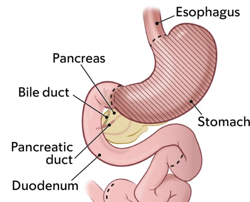
In a recent study, more than 90% of participants whose stomachs had been surgically removed to prevent cancer experienced a least one chronic complication 2 years out from their surgery. For some, the complications are life-altering.
Findings from a recent study will help clinicians counsel people who are considering preventive gastrectomy about the long-term impacts of the surgery.

Exploiting an ingenious combination of photochemical (i.e., light-induced) reactions and self-assembly processes, a team led by Prof. Alberto Credi of the University of Bologna has succeeded in inserting a filiform molecule into the cavity of a ring-shaped molecule, according to a high-energy geometry that is not possible at thermodynamic equilibrium. In other words, light makes it possible to create a molecular “fit” that would otherwise be inaccessible.
“We have shown that by administering light energy to an aqueous solution, a molecular self-assembly reaction can be prevented from reaching a thermodynamic minimum, resulting in a product distribution that does not correspond to that observed at equilibrium,” says Alberto Credi.
“Such a behavior, which is at the root of many functions in living organisms, is poorly explored in artificial molecules because it is very difficult to plan and observe. The simplicity and versatility of our approach, together with the fact that visible light—i.e., sunlight—is a clean and sustainable energy source, allow us to foresee developments in various areas of technology and medicine.”

Optical tweezers and related techniques provide extraordinary opportunities for research and applications in the physical, biological, and medical fields. However, certain requirements such as high-intensity laser beams, sophisticated electrode designs, additional electric sources, and low-conductive media, significantly impede their flexibility and adaptability, thus hindering their practical applications.
In a study published in The Innovation, a research team led by Dr. Du Xuemin from the Shenzhen Institutes of Advanced Technology (SIAT) of the Chinese Academy of Sciences reported a novel photopyroelectric tweezer (PPT) that combines the advantages of the light and electric fields. The PPT enables versatile manipulation in various working scenarios.
The proposed PPT consists of two key components, a near infrared (NIR) spectrum laser light source and a PPT device that includes a liquid medium and a photopyroelectric substrate.