A study in five volunteers undergoing surgery confirmed the existence of channels that may help drain waste from the brain.
Scientists have developed a nanoparticle-based treatment that successfully reversed Alzheimer’s disease in mice.
As detailed in a new paper published in the journal Signal Transduction and Targeted Therapy, the team co-led by the Institute for Bioengineering of Catalonia, Spain (IBEC), and West China Hospital, Sichuan University, developed bioactive “supramolecular drugs” that can proactively repair the blood-brain barrier.
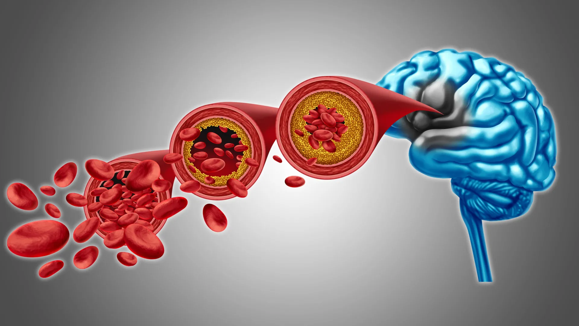
The barrier plays an important role in the health of the brain, defending it from harmful substances and other pathogens. Alzheimer’s has been linked to a weakening of the barrier’s integrity, allowing for impairing toxins to make it through.
Even small amounts of bisphenol A can lead to long-term health effects. When researchers studied adult rats exposed in the fetal stage, they found that females had developed a more masculine and males a more feminine gene expression pattern. This led to females progressing towards a cancer-like state, while males progressed towards metabolic syndrome, which can increase the risk of diabetes and heart disease.
Bisphenol A is a synthetic chemical with estrogen-like properties that is commonly used in food packaging materials. The substance is banned in many products, but is still present in some packaging. Levels of bisphenol A in people’s bodies are often above levels considered safe, with previous research showing that the substance can cause adverse health effects.
Females masculinized and males feminized In the current study, published in Communications Medicine, researchers investigated how bisphenol A affects the body during the fetal stage.
Chimeric Antigen Receptor (CAR) T cell therapies have revolutionized cancer treatment—but so far, their success has been largely limited to blood cancers. Solid tumors, which account for around 90% of all adult cancers, remain a major challenge because they are difficult for CAR T cells to infiltrate and are often highly heterogeneous, making them harder to target with a single therapy.
Researchers at Monash University, in collaboration with scientists from the Peter MacCallum Cancer Center, used CRISPR-based gene editing or a PTPN2 inhibitor to enhance the function of human CAR T cells engineered to recognize an antigen expressed on many solid tumors.
The study, led by Professor Tony Tiganis and Dr. Florian Wiede, was published in Science Translational Medicine.
Every cell in the body has the same DNA, but different cell types—such as muscle or brain cells—use different parts of it. Transcription factors help cells activate specific genes by reading certain DNA sequences, but since these sequences are common across the genome, scientists have long wondered how the factors know exactly where to bind.
Researchers in the Schübeler lab set out to address this question by looking at two closely related transcription factors—NGN2 and MyoD1—that steer cells toward becoming neurons and muscle cells, respectively. Using stem cells, they switched these transcription factors on one at a time and watched where they attached to the DNA and how they influenced gene expression. Their research is published in the journal Molecular Cell.
They found that the binding of transcription factors to the DNA molecule depends not only on the DNA sequence but also on how open the DNA is and which partner proteins are present. Sometimes, transcription factors act as “pioneer factors” and are able to open tightly packed DNA at specific sites to turn on genes. Small DNA changes—sometimes just one letter—and the proteins these factors partner with can affect whether genes are activated.
Using powerful 7-Tesla brain imaging, researchers mapped how the brainstem manages pain differently across the body. They discovered that distinct regions activate for facial versus limb pain, showing the brain’s built-in precision pain control system. The findings could lead to targeted, non-opioid treatments that use cannabinoid mechanisms instead of opioids, offering safer pain relief options.
“Over the last 10 or 15 years, scientists have really started to understand the fundamental underlying biology of the aging process. And they broke this down into 12 hallmarks of aging.”
Up next, Why 2025 is the single most pivotal year in our lifetime | Peter Leyden ► • Why 2025 is the single most pivotal year i…
We track age by the number of birthdays we’ve had, but scientists are arguing that our cells tell a different, more truthful story. Our biological age reveals how our bodies are actually aging, from our muscle strength to the condition of our DNA.
The gap between these two numbers may hold the key to treating aging – which could help save 100,000 lives per day and win us $38 trillion dollars.
00:00 Rethinking longevity.
01:27 Understanding aging.
02:58 Biological age and epigenetics.
04:29 New frontiers in longevity science.
08:04 Future possibilities and ethical questions.
10:24 The moral debate around living longer.
For the first time, a research team led by the Mark and Mary Stevens Neuroimaging and Informatics Institute (Stevens INI) at the Keck School of Medicine of USC has mapped the genetic architecture of a crucial part of the human brain known as the corpus callosum—the thick band of nerve fibers that connects the brain’s left and right hemispheres. The findings open new pathways for discoveries about mental illness, neurological disorders and other diseases related to defects in this part of the brain.
The corpus callosum is critical for nearly everything the brain does, from coordinating the movement of our limbs in sync to integrating sights and sounds, to higher-order thinking and decision-making. Abnormalities in its shape and size have long been linked to disorders such as ADHD, bipolar disorder, and Parkinson’s disease. Until now, the genetic underpinnings of this vital structure had remained largely unknown.
In the new study, published in Nature Communications, the team analyzed brain scans and genetic data from over 50,000 people, ranging from childhood to late adulthood, with the help of a new tool the team created that leverages artificial intelligence.
Fiber-optic technology revolutionized the telecommunications industry and may soon do the same for brain research.
A group of researchers from Washington University in St. Louis in both the McKelvey School of Engineering and WashU Medicine have created a new kind of fiber-optic device to manipulate neural activity deep in the brain. The device, called PRIME (Panoramically Reconfigurable IlluMinativE) fiber, delivers multi-site, reconfigurable optical stimulation through a single, hair-thin implant.
“By combining fiber-based techniques with optogenetics, we can achieve deep-brain stimulation at unprecedented scale,” said Song Hu, a professor of biomedical engineering at McKelvey Engineering, who collaborated with the laboratory of Adam Kepecs, a professor of neuroscience and of psychiatry at WashU Medicine.