Toggle light / dark theme

Gene editing tool reduces Huntington’s toxic protein fragments and symptoms in mice

A gene-editing tool designed to precisely rewrite the gene that causes Huntington’s disease reduced toxic protein fragments and symptoms associated with the disease in mice, researchers at the University of Illinois Urbana-Champaign report.

While other gene-based treatments have focused on turning the gene off, the Illinois team took a different approach. The researchers designed a base-editing tool to alter a specific point in the huntingtin gene so the cell’s machinery would skip over a small section prone to generating toxic fragments while preserving enough huntingtin protein to support its normal functions.

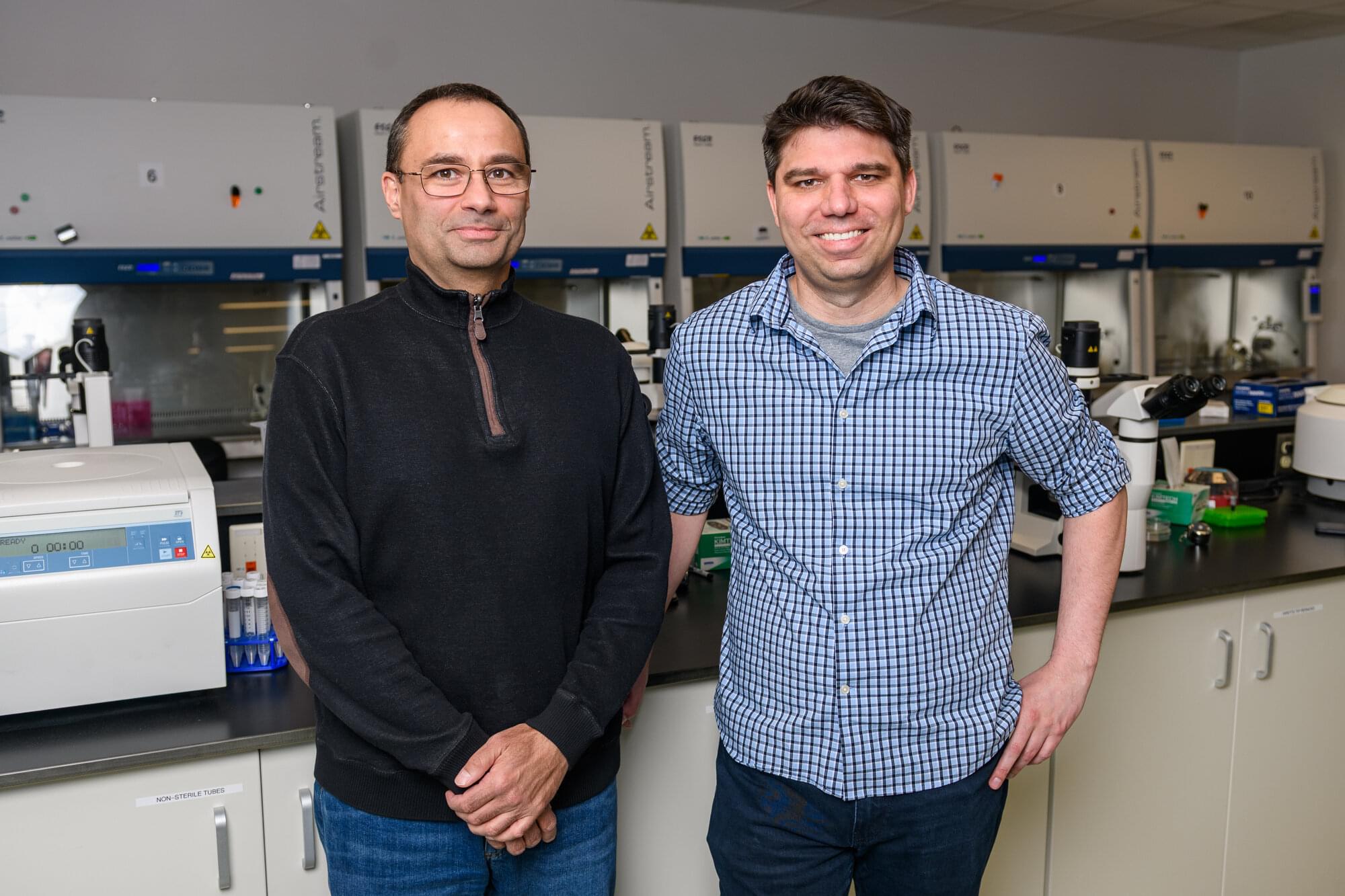
Led by Pablo Perez-Pinera and Thomas Gaj, professors of bioengineering at the U. of I., the researchers published their findings in the journal Nature Biomedical Engineering.
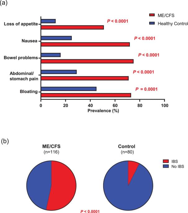
Gastrointestinal symptoms correlate with core clinical features and systemic inflammation in myalgic encephalomyelitis/chronic fatigue syndrome

Myalgic encephalomyelitis/chronic fatigue syndrome (ME/CFS) is a debilitating multisystem illness marked by fatigue, cognitive impairment, and post-exertional malaise. Gastrointestinal (GI) symptoms are frequently reported, yet their relationship to central features of the illness and biological correlates remains poorly understood.

We aimed to characterize GI symptom burden in ME/CFS and evaluate its associations with core clinical features and specific immune and inflammatory markers, with attention to potential gut-related contributions to disease expression.

GI symptoms and 49 additional symptoms across nine domains were assessed in 116 ME/CFS patients and 80 matched controls. Plasma C-reactive protein (CRP) and antibodies against dietary and microbial antigens were measured as indicators of systemic inflammation and putative gut-derived antigen exposure.

Defining Endogenous DMT Brain Biotypes: A Multi-Modal Neuroimaging Study

Could Your Brain Have Its Own “DMT Signature”? A New Research Proposal Aims to Find Out.

A new neuroscience research proposal is exploring a fascinating question: Do people naturally differ in their levels or activity of endogenous DMT, and could those differences be reflected in distinct brain “biotypes”?

Rather than administering DMT, the researchers propose analyzing an existing dataset of approximately 1,100 participants using multiple complementary measures, including:

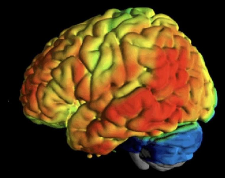
PET imaging to examine serotonin receptor systems.

Structural and functional MRI to assess brain anatomy and connectivity.

Diffusion MRI to evaluate white matter microstructure.

Blood biomarkers.

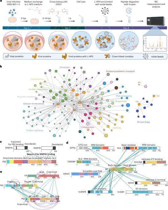
Mapping incell protein contact sites reveals hijacking of paraspeckles during influenza A virus infection Microbiology

In-cell cross-linking mass spectrometry maps protein contacts in influenza-infected human cells, revealing how the virus hijacks host membrane-trafficking factors and dismantles nuclear paraspeckles to enhance replication.

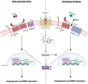
The calcium pump ATP2B1/PMCA1 regulates CNS vascular development by facilitating Norrin Frizzled4 signaling

Jo et al. identify the plasma membrane Ca2+−pump ATP2B1/PMCA1 as a regulator of endothelial Norrin/Frizzled4 and Wnt signaling in the CNS vasculature. Loss of ATP2B1 elevates intracellular Ca2+ and activates NFAT, suppressing β-catenin signaling and linking Ca2+ homeostasis to angiogenesis and blood-brain barrier integrity.

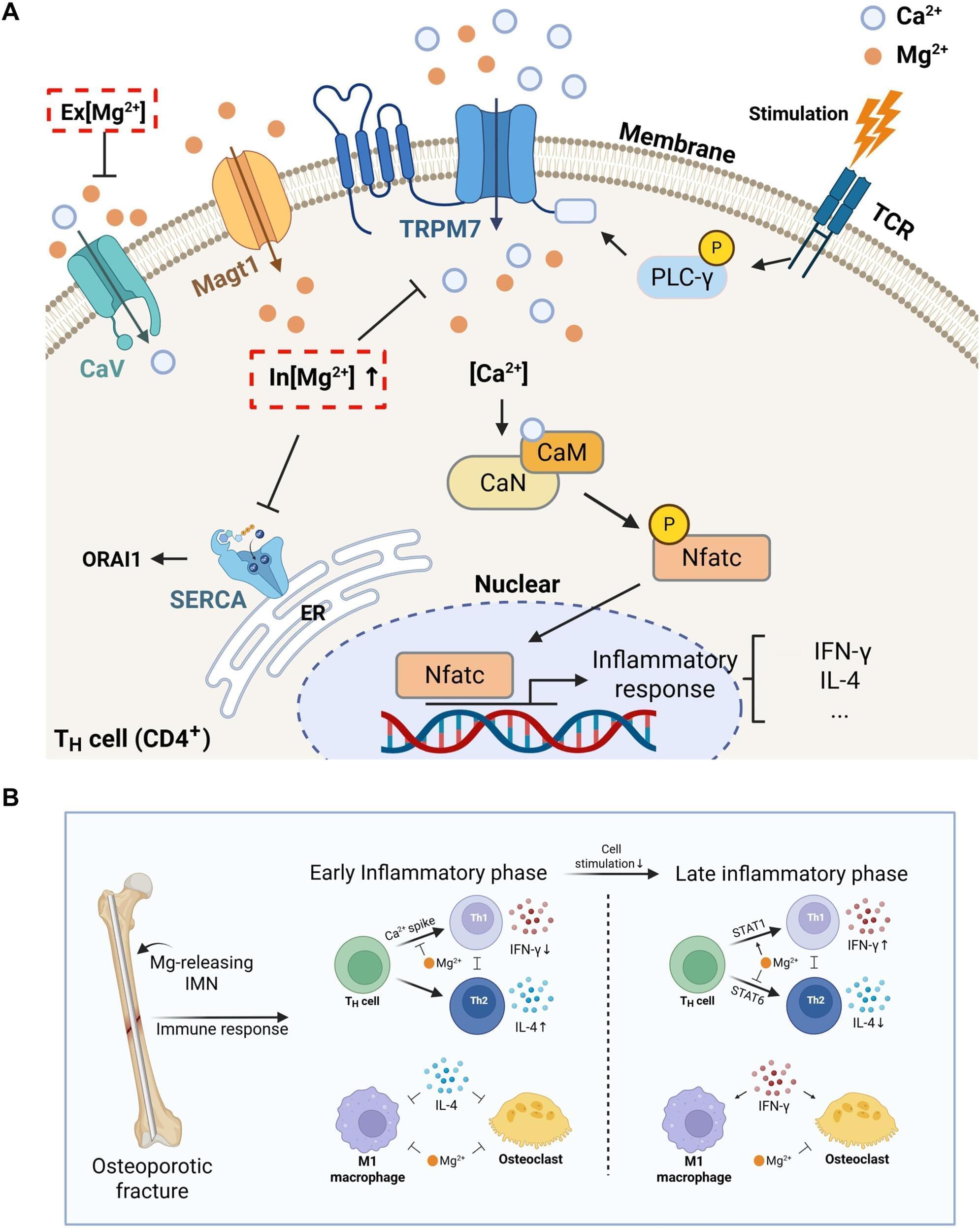
Bioceramic-coated implant improves osteoporotic fracture healing through timed magnesium release

Seoul National University (SNU) College of Engineering announced that a research team led by Nathaniel S. Hwang, a professor in the Department of Chemical and Biological Engineering, has developed a bioceramic fracture fixation material that promotes bone regeneration by precisely controlling the timing of magnesium ion (Mg²⁺) release to suppress inflammatory immune responses during osteoporotic fracture healing.

The research team discovered that magnesium ions do not always promote bone regeneration; rather, their effects on immune responses and bone healing vary depending on the timing and duration of release. Based on this finding, the team proposed a fracture fixation material that releases magnesium ions according to the stages of healing and demonstrated its bone regeneration efficacy through animal experiments.

Furthermore, the study suggests the possibility of advancing fracture treatment materials beyond simple mechanical fixation devices into therapeutic technologies that actively regulate immune responses according to healing stages. The newly developed material is expected to be applied to next-generation orthopedic medical devices and personalized bone regeneration therapies for patients with osteoporotic fractures.

Researchers train AI to detect diabetes and assign four diagnostic labels

Researchers developed a two-stage machine learning framework that detected diabetes and classified records as prediabetes, type 1, type 2, or type 3c diabetes using two public datasets. XGBoost showed strong internal performance, but inconsistent model rankings, non-standard variables, and the absence of external clinical validation limit immediate use.

New mechanism reveals how heart cells resist regenerative reprogramming

Specialized cells in the human body make biological trade-offs to perform certain jobs. To support the large, hardworking hearts that power other organs, heart cells have evolved to be extremely efficient and resilient. Because the adult human heart cannot repair itself the way skin does, scientists have studied ways to “reprogram” heart cells to regenerate after heart attacks. Heart cells have very stable identities, however, and are resistant to reprogramming.

Scientists at Sanford Burnham Prebys Medical Discovery Institute and collaborators at Johns Hopkins University School of Medicine published findings in Nature Communications that cover a new mechanism cells use to protect their identities and ward off reprogramming attempts. Continued study of the obstacles to cellular reprogramming may lead to treatments that can help the heart repair itself after injury.

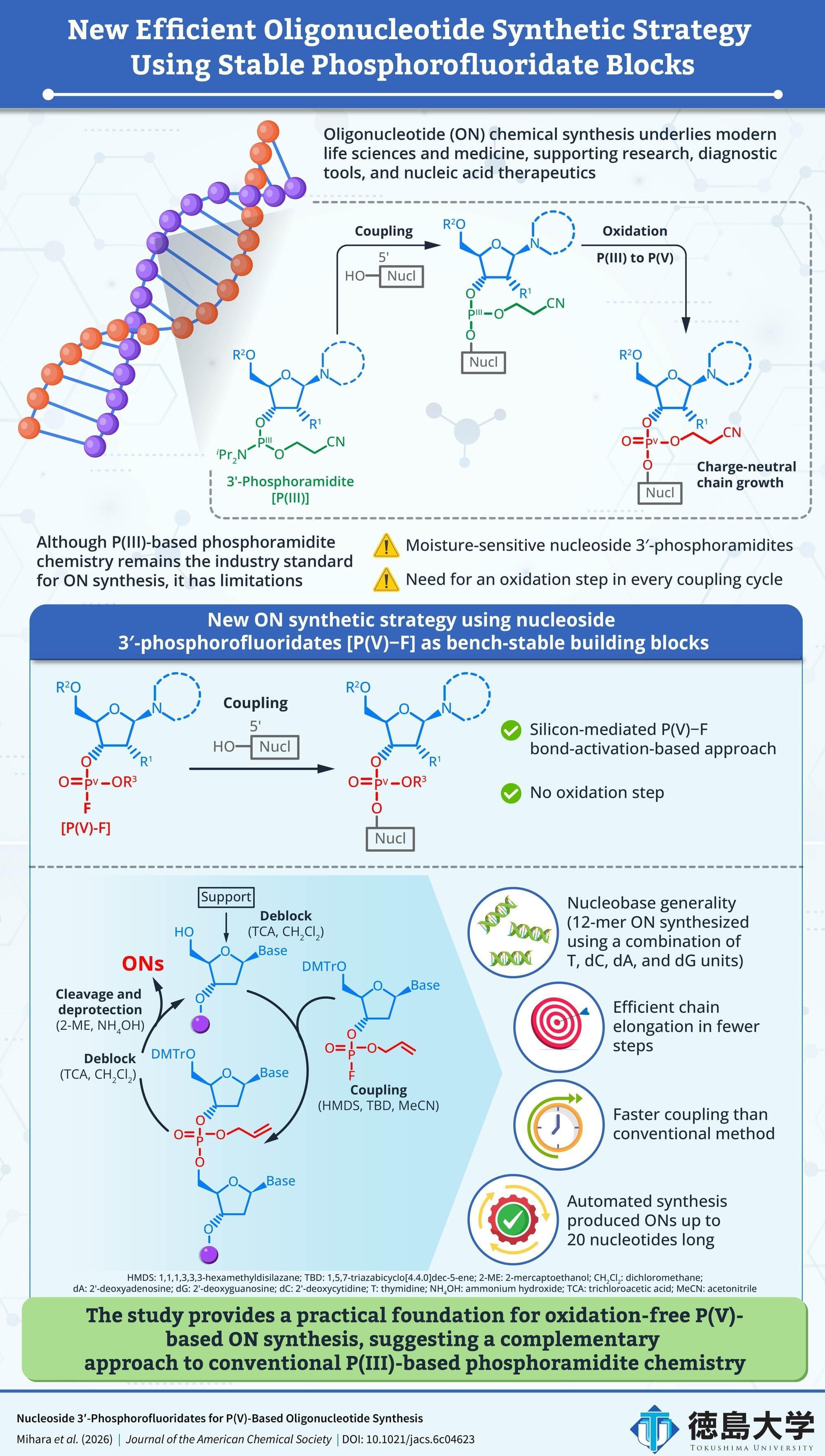
Stable DNA building blocks enable faster oligonucleotide synthesis without oxidation

The chemical synthesis of oligonucleotides (ONs) is central to modern molecular biology, diagnostics and nucleic acid therapeutics. While demand for high-quality ONs is increasing, the conventional synthetic method has long-standing efficiency challenges. Widely adopted P(III)-phosphoramidite-based ON synthesis requires an oxidation step after every nucleotide coupling cycle and uses moisture-sensitive building blocks, adding complexity to the workflow and slowing the process.

Early studies of ON synthesis showed that pentavalent phosphorus [P(V)] chemistry could form linkages between nucleotides. However, practical limitations, including unstable intermediates, slow coupling, harsh deprotection or poor performance during chain elongation, prevented these methods from replacing P(III)-based phosphoramidite chemistry.

A recent study led by Associate Professor Noriko Saito-Tarashima of the Graduate School of Pharmaceutical Sciences at Tokushima University in Japan, along with Nana Mihara, a doctoral student from the same institution, investigated whether nucleoside 3′-phosphorofluoridates [P(V)–F] could be used as stable building blocks for ON synthesis without requiring a separate oxidation step.

/* */