Toggle light / dark theme

Intestine, Liver, Kidney Proximal Tubule, Blood-Brain Barrier and Skeletal Muscle

This is definitely a share that is interesting to many studying synthetic organs and their acceptance into the human body as well as the work occurring on Quantum biology as well.


The goal of in vitro and in vivo toxicity testing is to identify compounds that would predict adverse reactions in humans. Olson et al. found that only 70% of human toxicity was predicted from animal testing. Currently we rely on traditional toxicity testing in animals, a 1930’s methodology that is now challenged due to questionable relevance to human risk, high cost, ethical concerns, and throughput that is too limited for the nearly 80,000 industrial chemicals not yet tested for safety. Additionally, testing usually extrapolates acute, high dose animal results to chronic, low dose human exposures, thereby risking rejection or limiting the use of drugs, industrial chemicals or consumer products. Moreover, the ability of lab animal target organ toxicity to predict dose-limiting toxicity in the corresponding human organ varies widely, from a low of 30% for human cutaneous toxicity, to 50–60% for human hepatotoxicity, to a high of 90% for hematological drug toxicity. Animal drug efficacy models are also notoriously discordant. In an analysis of six drugs to treat head injury, hemorrhage, acute ischemic stroke, neonatal respiratory distress syndrome, and osteoporosis, it was found that efficacy was similar in animals and humans for three drugs but was dissimilar for another three. In oncology drug development, animal models often over-predict anti-tumor efficacy in humans3,4. Examples such as these highlight the need to continue research into methods that reduce the dependence on laboratory animals for toxicity testing of environmental chemicals, determine efficacy and toxicity in drug development, serve as a mimic of human diseases, and provide patient-specific guidance in the emerging field of precision medicine.

Recent advances in bioengineered materials, microfluidic technology, and the availability of human primary, immortalized, and induced pluripotent stem cell (iPSC)-derived cells are enabling development of human microphysiological systems (MPS), sometimes called “organs-on-a-chip” or “human-on-a-chip,” that use multiple organ-specific human cells to recapitulate many functional and structural properties of a human organ. It is now generally accepted and supported by data that cellular responses to drugs in most human organs are more accurately approximated in 3D cell cultures than in traditional static 2D cell cultures5,6. Microfluidic perfusion further improves model performance by providing a flow of nutrients and oxygen and the removal of waste products from the cell cultures. Physiologically relevant flow increases oxygen consumption, Krebs cycle activity and secretion of synthesized proteins, and decreases expression of the hypoxia HIF1 gene. Flow also improves the absorption and metabolism of compounds like benzo[a]pyrene6,8,9. The large number of recent publications reviewing organ MPS models indicates a high degree of interest by industrial and academic researchers, granting agencies and other stakeholders10,11,12,13. In addition to the stand-alone MPS, investigators are linking MPS to study organ-organ functional interactions, efficacy, PK and toxicology14,15,16,17,18.

An obvious approach to linking organs is direct coupling of the media stream outflow from one organ into the inflow of the next by use of tubing or a connecting channel. Some limitations to this approach include the requirement for a common medium, difficulty in reducing metabolic wastes to the next organ, organ-specific flow rates and adequate oxygenation of all modules in the system19. These requirements are most easily addressed when the linked organ modules are designed and developed at the same time and in the same laboratory, but even when the organ modules are co-developed, the proper scaling between organ modules is a significant design and calculation challenge. Although organ modules can be sized using allometric scaling20, the resulting functional capacity of the individual organ models may not scale the same.

Faraday Rotation Spectroscopy for Speedy Medical Testing

Nice.


Researchers at the University of Central Florida (UCF) in the US are combining nanoscience with the principle of Faraday rotation, a magnetic phenomenon discovered in 1845, in a new method for speedy medical tests.

The team applied the magneto-optical technique, called frequency-domain Faraday rotation spectroscopy—or fd-FRS, to characterize proteins, using antibody-functionalized magnetic nanoparticles (MNPs).

“Fd-FRS is a new way of looking at the interaction between light and magnetized material,” says Shawn Putnam, assistant professor UFC’s College of Engineering & Computer Science. “We covered the surfaces of magnetic nanoparticles with a layer of biological sensors and measured their movement after exposure to their corresponding biological targets for detection.”

The Chan Zuckerberg Biohub invests $50 million in its first 47 research initiatives

The Chan Zuckerberg Biohub, a non-profit medical research organization started by Priscilla Chan and Mark Zuckerberg (and not to be confused with the Chan Zuckerberg Initiative, a limited liability corporation to advance human potential) today announced it would be doling out a total of $50 million to its first cohort of disease investigators.

The Biohub brings together Bay Area universities including the University of California, Berkeley; the University of California, San Francisco; and Stanford. One of the keystones to this collaboration effort is the investigative program.

Exes Don Johnson & Melanie Griffith’s Bizarre Medical Experiments — To Live Longer!

Forgive the source, but i saw this in the grocery store tabloid, still thought it was kind of interesting. These are the kind of people we should be trying to reach out to for life extension tech funding; their jobs are their looks, they have lots of money, and probably a million and one connections.


Agingexes Melanie Griffith and Don Johnson reunited at a mysterious German clinic with ties to a controversial doctor specializing in “fountain of youth” cures — including stem cell therapy!

In a bombshell exclusive, Radar caught the couple making a hush-hush visit to the ultra-private ACQUA Klinik in Leipzig — and Don’s wife was nowhere in sight!

There, they met with Dr. Augustinus Bader, a professor of stem cell technology who’s reportedly developed a “miracle” cure for aging. The visit also ignited speculation Melanie, 59, and Don, 67, may be rekindling their relationship — for the third time!

What is the difference between DNA and RNA

To understand 1st how to solve things like cancer, or other immune deficiency disorders, or to solve the human system’s other mysteries; one must understand DNA v. RNA.


DNA is a deoxyribonucleic acid and is a hereditary material in all living organism. It is located in the nucleus of cell which is known as nuclear DNA. But small amount of DNA is also found in the mitochondria known as mtDNA or mitochondrial DNA whereas RNA is ribonucleic acid present in all living cells. It carries instructions from DNA which controls the synthesis of proteins but in some viruses RNA, instead of DNA carries the genetic information. Do you know who had reported first time nucleic acids in 1871 — Friedrich Miescher from nuclei of pus cells.

DNA deoxyribonucleic

Source: www.static.diffen.com

How Genetics define the hereditary characteristics of human?

Researchers Explain Mechanism Controlling Cell Protein Generation

Yesterday, I shared the new cancer research discovery by Prof. Samie Jaffrey and his team. The following provides additional details on their discovery.


The human body is made of trillions of cells. Each fulfills a specific purpose, undergoes tremendous wear and tear and is eventually replaced. Despite extensive research, some questions related to protein production that fuels this simple process have gone unanswered. Fascinated by these intricacies, Prof. Samie Jaffrey, pharmacology, may have found part of the answer.

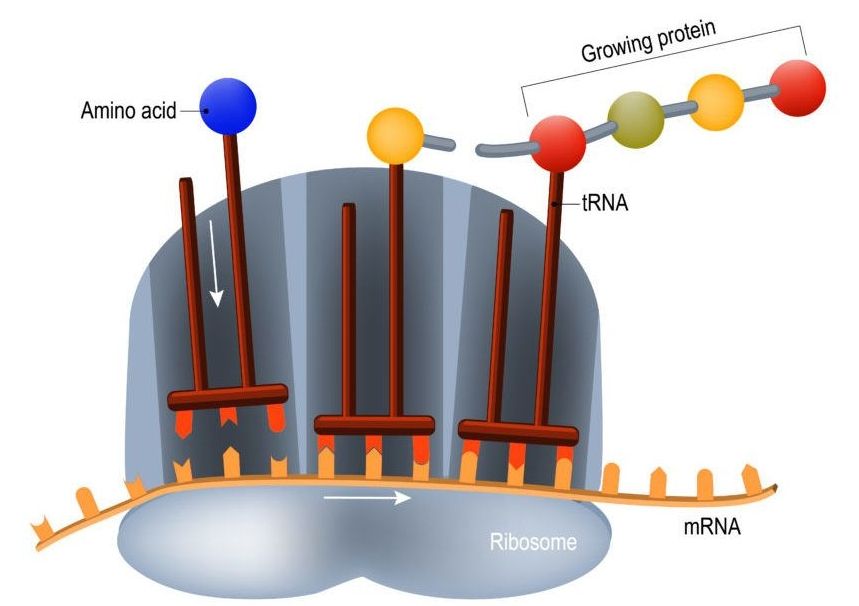
Jaffrey and his team discovered that messenger RNA molecules, responsible for conveying genetic information to protein producers in the cell, have special features that predetermine how much protein they generate. The discovery could provide scientists with a greater understanding of the causes of cancer and ways of regulating it.

“Over the past few years we have found that methylation [deposition of methyl groups] inside the mRNA can have effects on translation [the process of translating genetic information to physical protein production] and stability,” Jaffrey said.

Genetics Is Giving Way to a New Science of Life

By Jonathan Latham, PhD

Test your understanding of the living world with this simple question. What kind of biomolecule is found in all living organisms? If your answer is “DNA”, you are incorrect. The mistake is very forgiveable though. The standard English-language biology education casts DNA (DeoxyriboNucleic Acid) as the master molecule of life, coordinating and controlling most, if not all, living functions. This master molecule concept is popular. It is plausible. It is taught in every university and high school. But it is wrong. DNA is no master controller, nor is it even at the centre of biology. Instead, science overwhelmingly shows that life is self-organised and thus the pieces are in place for biology to undergo the ultimate paradigm shift.

/* */