Toggle light / dark theme

Study shows that dendritic plasticity contributes to the integration of memories

Past neuroscience studies suggest that memories of events that occurred at short time intervals from one another are often connected, via a process referred to as memory linking. While memory linking is now a well-documented phenomenon, its neural underpinnings have not been fully elucidated.

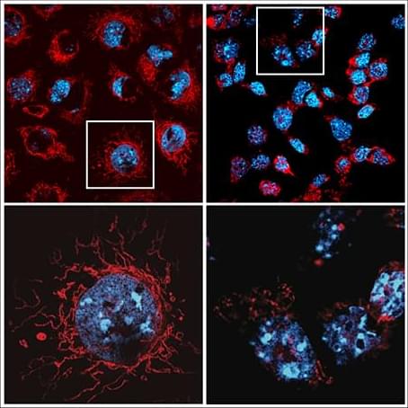
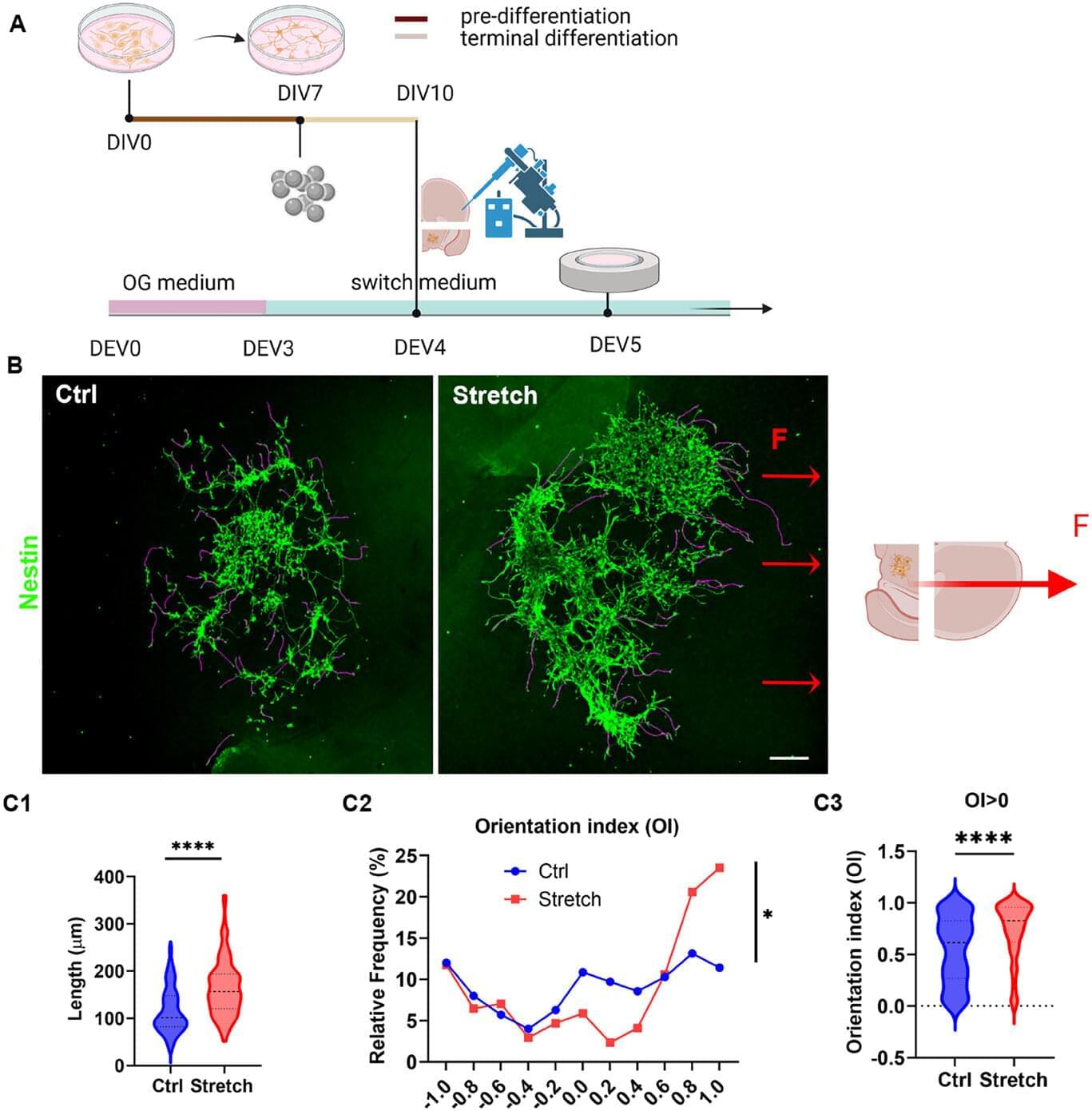
Researchers at the University of California Los Angeles (UCLA) recently carried out a study aimed at better understanding the neural processes that contribute to memory linking in the . Their findings, published in Nature Neuroscience, suggest that dendritic plasticity, the adaptation of dendrites (i.e., branch-like extensions of neurons) over time, plays a key role in the linking of memories.

“A few years back, in a landmark study published in Nature in 2016, we demonstrated that memories formed a few hours apart are linked because they are stored in a common set of neurons in the hippocampus,” Alcino Silva, senior author of the paper, told Medical Xpress. “We wanted to know: Where within these neurons are these memories stored and linked? What was causing these neurons to be recruited?”

Algorithm sheds light on ‘disordered’ proteins once considered too difficult to study

Intrinsically disordered proteins (IDPs) do not attain a stable secondary or tertiary structure and rapidly change their conformation, making structure prediction particularly challenging. Although these proteins exhibit chaotic and “disordered” structures, they still perform essential functions.

IDPs comprise approximately 30% of the and play important functional roles in transcription, translation, and signaling. Many mutations linked to , including (ALS), are located in intrinsically disordered protein regions (IDRs).

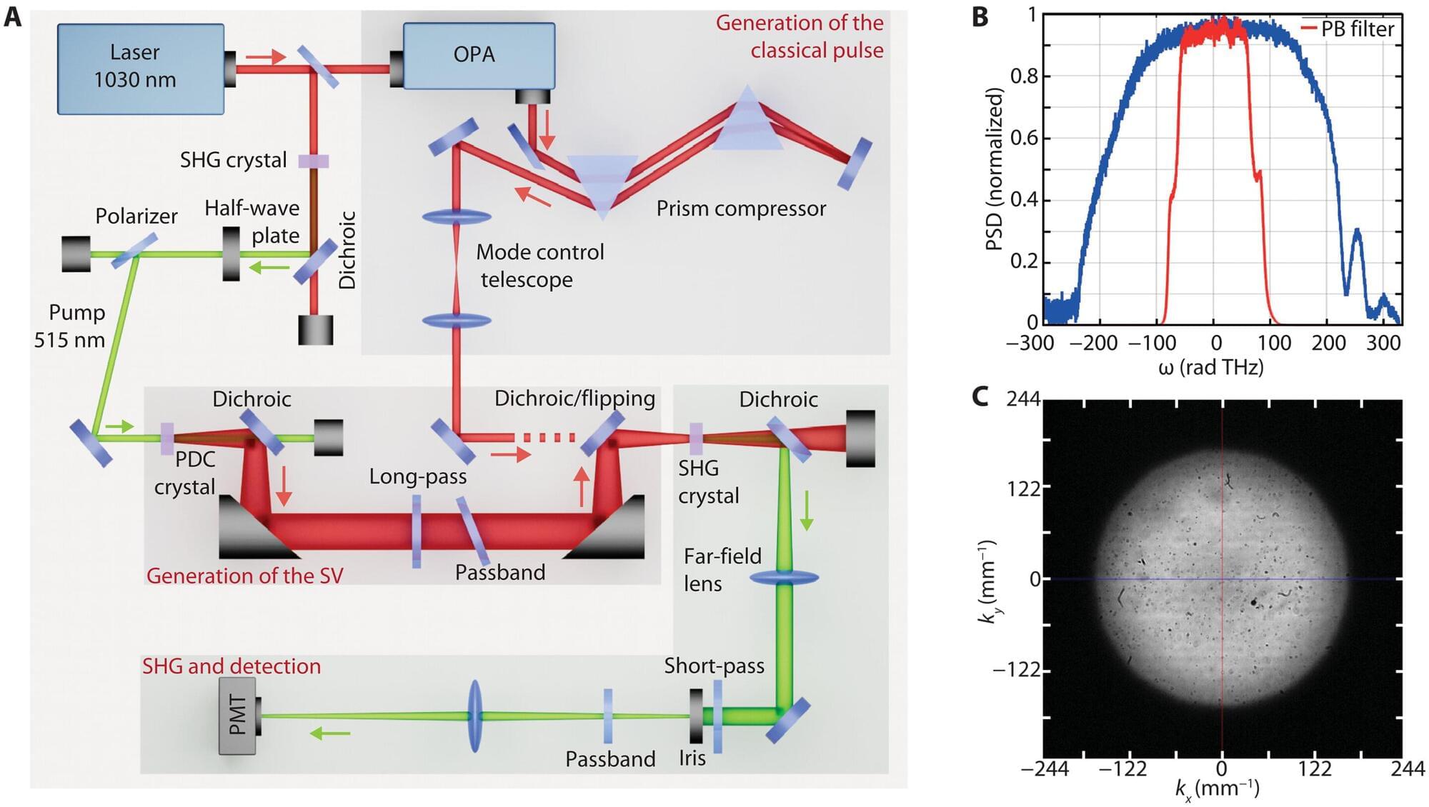
Powerful machine-learning algorithms, including AlphaFold and RoseTTAFold, cannot provide realistic representations of these ‘disordered’ and ‘chaotic’ protein regions as a whole. This is because they have not been trained on such data and because these proteins exhibit inherent dynamic behavior, adopting a range of conformations rather than a single stable one.

Heat-activated receptor could unlock new pain therapies

Touch a hot plate and your hand flies back. While the response is almost instant, researchers are still working to better understand the molecular mechanisms behind these sensations of heat and pain.

Now, investigators at the Jacobs School of Medicine and Biomedical Sciences at the University at Buffalo have uncovered how heat causes a critical receptor protein within cells to unfold and relay pain. This newfound activation mechanism could open up new therapeutic targets for treating pain and contribute to the development of needed alternatives to opioids.

The study is published in Proceedings of the National Academies of Sciences.

Entangled polymers and nanosheets create skin-like, self-healing hydrogel

We all encounter gels in daily life—from the soft, sticky substances you put in your hair to the jelly-like components in various foodstuffs. While human skin shares gel-like characteristics, it has unique qualities that are very hard to replicate. It combines high stiffness with flexibility, and it has remarkable self-healing capabilities, often healing completely within 24 hours of an injury.

Until now, artificial gels have either managed to replicate this high stiffness or natural skin’s self-healing properties, but not both. Now, a team of researchers from Aalto University and the University of Bayreuth are the first to develop a hydrogel with a unique structure that overcomes earlier limitations, opening the door to applications such as , , soft robotics sensors and artificial skin.

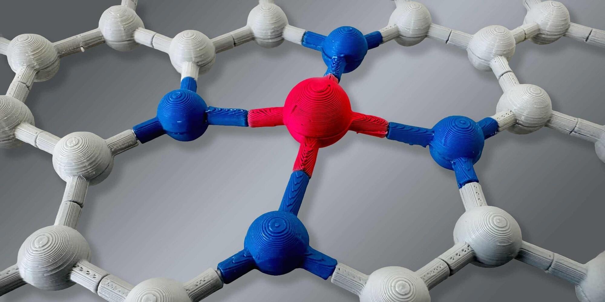
In the study, the researchers added exceptionally large and ultra-thin specific clay nanosheets to hydrogels, which are typically soft and squishy. The result is a highly ordered structure with densely entangled polymers between nanosheets, not only improving the mechanical properties of the hydrogel but also allowing the material to self-heal.

Innovative biorobotic arm uses artificial muscles to combat tremors, paving way for wearable solutions

It is estimated that about 80 million people worldwide live with a tremor. For example, those who live with Parkinson’s disease. The involuntary periodic movements sometimes strongly affect how patients are able to perform daily activities, such as drinking from a glass or writing.

Wearable soft robotic devices offer a potential solution to suppress such tremors. However, existing prototypes are not yet sophisticated enough to provide a real remedy.

Scientists at the Max Planck Institute for Intelligent Systems (MPI-IS), the University of Tübingen, and the University of Stuttgart under the Bionic Intelligence Tübingen Stuttgart (BITS) collaboration want to change this. The team equipped a biorobotic arm with two strands of strapped along the forearm.

Feeling is believing: Bionic hand ‘knows’ what it’s touching, grasps like a human

Johns Hopkins University engineers have developed a pioneering prosthetic hand that can grip plush toys, water bottles, and other everyday objects like a human, carefully conforming and adjusting its grasp to avoid damaging or mishandling whatever it holds.

The system’s hybrid design is a first for robotic hands, which have typically been too rigid or too soft to replicate a human’s touch when handling objects of varying textures and materials. The innovation offers a promising solution for people with hand loss and could improve how robotic arms interact with their environment.

Details about the device appear in Science Advances.

Gut Microbe Composition During Infancy May Protect Against Diabetes in Later Life

Scientists headed by a team at the University of Utah Health have reported on research in mice suggesting that microbiome composition during infancy can shape development of pancreatic insulin-producing cells, leading to long-term changes in metabolism and impacting on diabetes risk later in life. The study, reported in Science by research co-lead June Round, PhD, professor of pathology at University of Utah Health, and colleagues, identified what the team describes as “a critical neonatal window in mice when microbiota disruption results in lifelong metabolic consequences stemming from reduced β cell development.”

Round suggests that understanding how the microbiome impacts metabolism could potentially lead to microbe-based treatments to prevent type 1 diabetes. “What I hope will eventually happen is that we’re going to identify these important microbes, and we’ll be able to give them to infants so that we can perhaps prevent this disease from happening altogether.”

In their published paper, titled “Neonatal fungi promote lifelong metabolic health through macrophage-dependent β cell development,” the team concluded that their results “… identify fungi as critical early-life commensals that promote long-term metabolic health …”

/* */