Vold et al. studied two SNAP-25 variants with different clinical severity. Variants destabilize the SNARE complex and reduce binding to the Munc18-1:VAMP2:syntaxin-1 acceptor complex, with correlated effects on neurotransmitter release. Effects of co-expression of variant and wild-type SNAP-25 were modeled by assuming the co-existence of both species in a ring of SNARE complexes.
Category: biotech/medical – Page 283

Using synthetic biology and AI to address global antimicrobial resistance threat
James J. Collins, the Termeer Professor of Medical Engineering and Science at MIT and faculty co-lead of the Abdul Latif Jameel Clinic for Machine Learning in Health, is embarking on a multidisciplinary research project that applies synthetic biology and generative artificial intelligence to the growing global threat of antimicrobial resistance (AMR).
The research project is sponsored by Jameel Research, part of the Abdul Latif Jameel International network. The initial three-year, $3 million research project in MIT’s Department of Biological Engineering and Institute of Medical Engineering and Science focuses on developing and validating programmable antibacterials against key pathogens.
AMR — driven by the overuse and misuse of antibiotics — has accelerated the rise of drug-resistant infections, while the development of new antibacterial tools has slowed. The impact is felt worldwide, especially in low-and middle-income countries, where limited diagnostic infrastructure causes delays or ineffective treatment.
Digital twin reveals how eye cells lose their organization in leading cause of vision loss
National Institutes of Health (NIH) researchers have developed a digital replica of crucial eye cells, providing a new tool for studying how the cells organize themselves when they are healthy and affected by diseases. The platform opens a new door for therapeutic discovery for blinding diseases such as age-related macular degeneration (AMD), a leading cause of vision loss in people over 50. The study is published in the journal npj Artificial Intelligence.
“This work represents the first-ever subcellular resolution digital twin of a differentiated human primary cell, demonstrating how the eye is an ideal proving ground for developing methods that could be used more generally in biomedical research,” Kapil Bharti, Ph.D., scientific director at the NIH’s National Eye Institute (NEI).
The researchers created a highly detailed, 3D data-driven digital twin of retinal pigment epithelial (RPE) cells, which perform vital recycling and supportive roles to light-sensing photoreceptors in the retina. In diseases such as AMD, RPE cells die, which eventually leads to the death of photoreceptor cells, causing loss of vision.

Cell division spindles self-organize like active liquid crystals—a theory that holds up
When a cell divides, it performs a feat of microscopic choreography—duplicating its DNA and depositing it into two new cells. The spindle is the machinery behind that process: It latches onto chromosomes (where DNA is stored) and separates them so they can settle into their new homes. This tricky process can sometimes go wrong, causing infertility, genetic disorders, or cancer.
Scientists have a good understanding of what spindles are made of: long, thin rods called microtubules as well as a variety of associated motor proteins. However, how these microtubules interact and organize to guide the spindles’ function has remained a mystery.
One approach to understand how the spindle self-organizes is to treat it like an active liquid crystal. Liquid crystals, like spindles, are made up of elongated subunits. Unlike liquid crystals in LCD displays, which require an external electric field to reorient their subunits, spindles are active materials that generate forces internally.
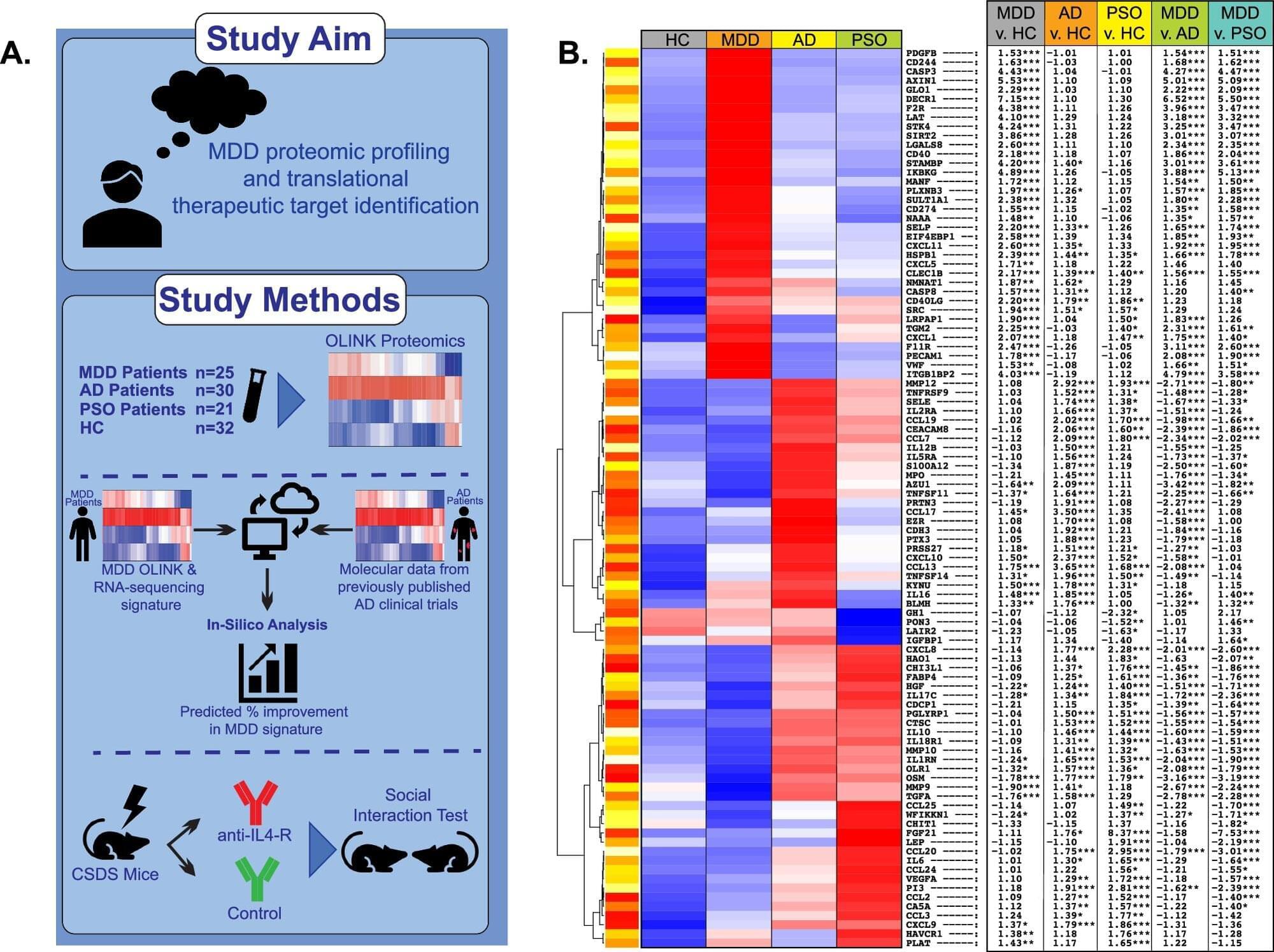
Major depressive disorder shares immune abnormalities and potential therapies with inflammatory skin diseases
A team of leading clinical research scientists from the Departments of Psychiatry and Dermatology at the Icahn School of Medicine at Mount Sinai has found that the serum of patients with major depressive disorder shares immune abnormalities with inflammatory skin diseases, most notably the common Th2 immune pathway that is implicated in atopic dermatitis. Because these skin diseases are treatable, the findings suggest new therapeutic possibilities for psychiatric illness as well.
The study findings, published in Molecular Psychiatry, underscore the potential role of the Th2 axis in major depressive disorder and highlight the potential of targeting this specific immune pathway that involves interleukin-4 receptor alpha, a cell receptor known to play a key role in regulating inflammation, as a disease-modifying treatment for this psychiatric disorder.
Furthermore, the back-translational drug repurposing strategy employed in this study may offer a new approach to identifying immunomodulatory drugs in psychiatry.
Rocket science? 3D printing soft matter in zero gravity
What happens to soft matter when gravity disappears? To answer this, UvA physicists launched a fluid dynamics experiment on a sounding rocket. The suborbital rocket reached an altitude of 267 km before falling back to Earth, providing six minutes of weightlessness.
In these six minutes, the researchers 3D-printed large droplets of a soft material similar to the inks used for bioprinting —a developing technology that shows huge potential for regenerative and personalized medicine, tissue engineering and cosmetics. Bioprinting involves 3D-printing a mix of cells and bio-inks or bio-materials in a desired shape, often to construct living tissues.
The experiment was called COLORS (COmplex fluids in LOw gravity: directly observing Residual Stresses). Using a special optical set-up, the researchers could see where the printed material experienced internal stresses (forces) as the droplets spread and merged. Stressed regions stand out as bright colors in the experiment. Investigating how and where these stresses emerge is important because they can get frozen in a material as it solidifies, creating weak points where 3D-printed objects are most likely to break.
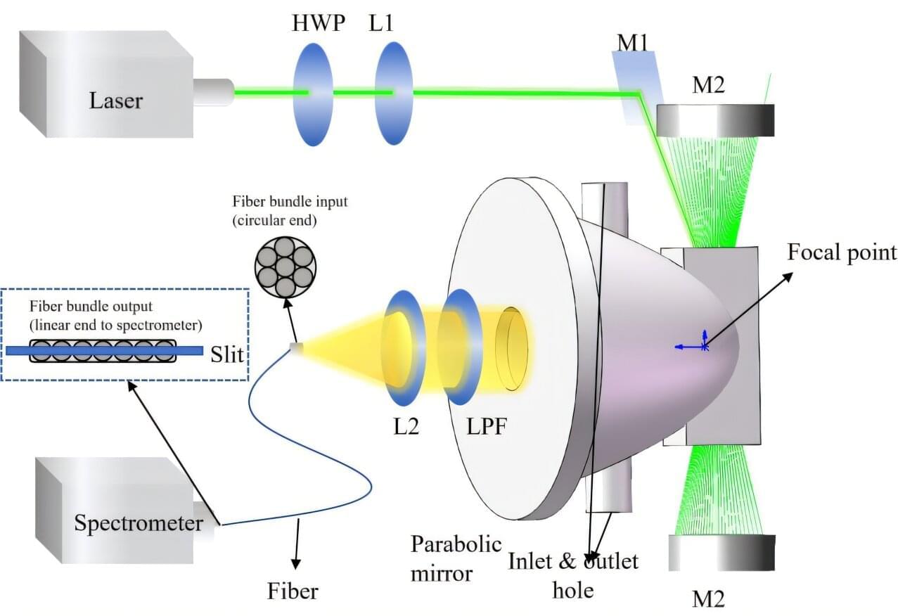
Parabolic mirror-enhanced Raman spectroscopy enables high-sensitivity trace gas detection
A research team led by Prof. Fang Yonghua from the Hefei Institutes of Physical Science of the Chinese Academy of Sciences proposed and systematically optimized a novel parabolic mirror cavity-enhanced Raman spectroscopy (PMCERS) technique, achieving a marked improvement in gas detection sensitivity through the integration of advanced optical design and signal processing methods. These results were published in Optics & Laser Technology.
Multi-component gas detection is important for environmental, industrial, and medical applications. Raman spectroscopy is well-suited for this purpose because it enables the simultaneous, water-vapor-free detection of multiple gas species. However, its inherently weak scattering limits sensitivity. Conventional cavity-enhanced approaches relying on lens-based collection have a limited numerical aperture, resulting in inefficient capture of three-dimensionally distributed Raman signals.
In this study, the team developed a parabolic mirror-based cavity-enhanced Raman spectroscopy system that leverages the large-aperture characteristics of parabolic mirrors to significantly improve Raman signal collection. Through the systematic optimization of the cavity structure, an efficient closed-loop optical path was established, effectively eliminating signal collection blind spots and suppressing stray-light interference.
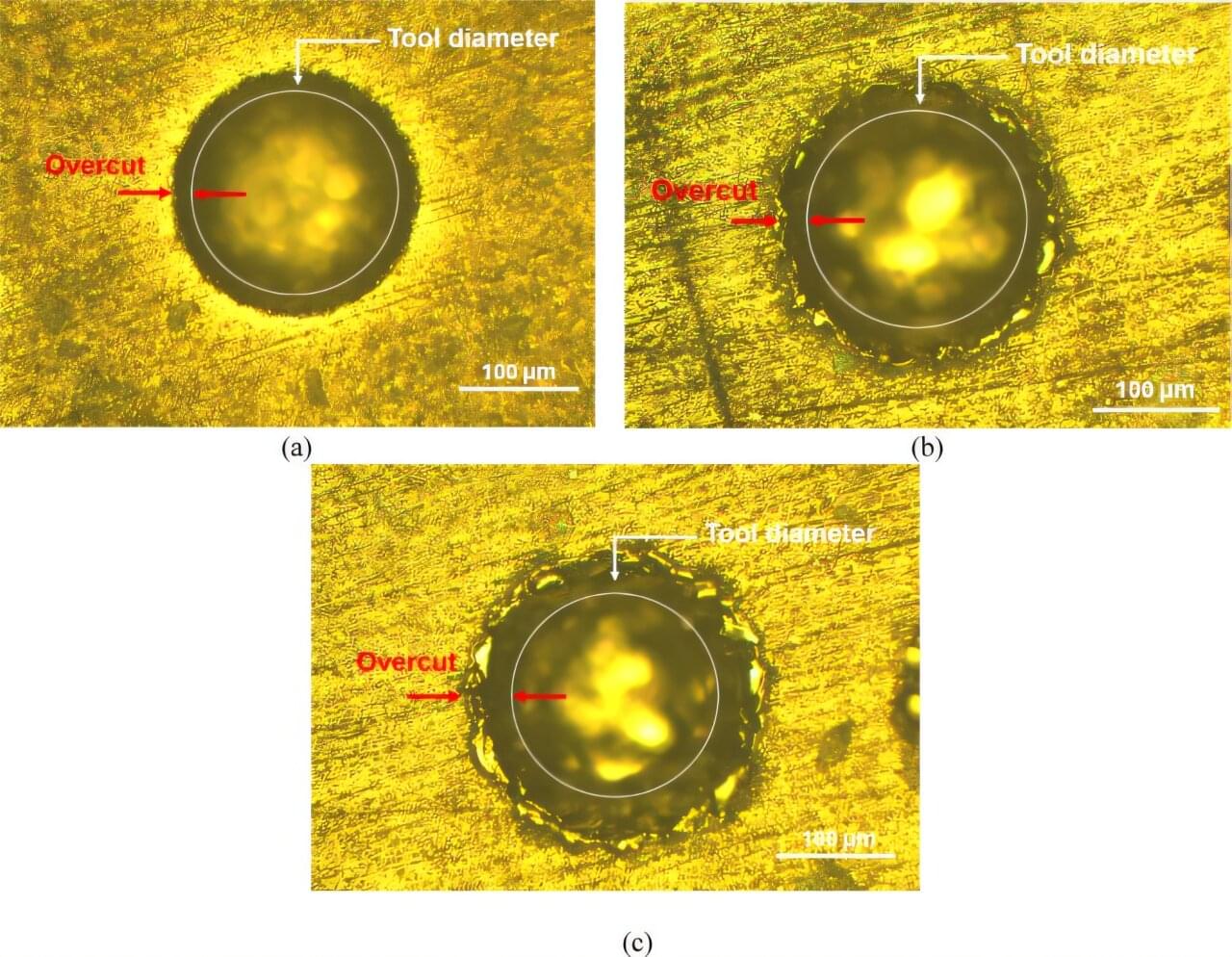
AI-guided micromachining advances next-generation biocompatible titanium alloys
Researchers have developed a new machine-learning-assisted approach to optimize micro-electro-discharge machining (µ-EDM) of a next-generation biocompatible titanium alloy, potentially improving the manufacturing of advanced medical and aerospace components.
The work is published in the journal Scientific Reports.
Titanium alloys are widely used in biomedical implants, aerospace systems, and automotive engineering due to their strength, corrosion resistance, and low weight. However, the commonly used alloy Ti–6Al–4V contains aluminum and vanadium, elements associated with long-term toxicity risks in biomedical applications.