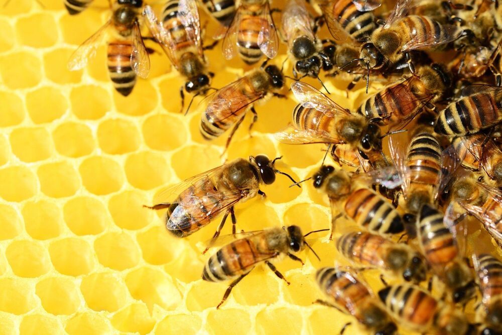
Toggle light / dark theme

Scientists Print Functional Human “Mini-Livers”

A team of Brazilian researchers have succesfully bioprinted tiny organoids that perform all of the human liver’s functions, Brazilian news service Agência FAPESP reports — functions including building proteins, storing vitamins and secreting bile.

The researchers had to cultivate and reprogram human stem cells, and then 3D print them in layers to form tissue.

While the “mini-livers” perform the functions of a liver, they’re unfortunately still a far cry from an actual full-scale liver.

Keep exercising: New study finds it’s good for your brain’s gray matter

Cardiorespiratory exercise—walking briskly, running, biking and just about any other exercise that gets your heart pumping—is good for your body, but can it also slow cognitive changes in your brain?

A study in Mayo Clinic Proceedings from the German Center for Neurodegenerative Diseases provides new evidence of an association between and brain health, particularly in and total brain volume—regions of the brain involved with cognitive decline and aging.

Brain tissue is made up of gray matter and filaments called white matter that extend from the gray matter cells. The volume of gray matter appears to correlate with various skills and cognitive abilities. The researchers found that increases in peak oxygen uptake are strongly associated with increased gray matter volume.

A new way to warm up frozen tissue could help with the organ shortage

This technology may one day be used to revive patient suspended in cryonics.


A new way to warm up frozen tissue using tiny vibrating particles could one day help with the problem of organ shortages.

We know how to cool organs to cryogenic temperatures, which is usually below 320 degrees Fahrenheit. But the organs can’t be stored for long — sometimes only four hours for heart and lungs — because they get damaged when you try to warm them up. As a result, more than 60 percent of donor hearts and lungs aren’t transplanted. In a study published today in Science Translational Medicine, scientists used nanoparticles to warm up frozen tissue quickly and without damaging the organs. Within a decade, this could lead to being able to store entire organs in organ banks for a long period of time, the authors say.

For today’s study, the team rewarmed 50 milliliters of tissue and solution with magnetic nanoparticles. Magnetic particles create heat in electromagnetic fields, says study co-author Zhe Gao, an post-doc studying nanotechnology at the University of Minnesota. Basically, the scientists infused a tissue with a special kind of nanoparticle made of silica-coated iron oxide. Then, they expose it to a magnetic field. Think of the nanoparticles as antennae. Once they get pick up the “signal” from the magnetic fields, they start to vibrate, and this creates the heat that warms up the organ quickly.

Decoder translates brain activity into speech

Neurological conditions or injuries that result in the inability to communicate can be devastating. Patients with such speech loss often rely on alternative communication devices that use brain–computer interfaces (BCIs) or nonverbal head or eye movements to control a cursor to spell out words. While these systems can enhance quality-of-life, they can only produce around 5–10 words per minute, far slower than the natural rate of human speech.

Researchers from the University of California San Francisco today published details of a neural decoder that can transform brain activity into intelligible synthesized speech at the rate of a fluent speaker (Nature 10.1038/s41586-019‑1119-1).

“It has been a longstanding goal of our lab to create technology to restore communication for patients with severe speech disabilities,” explains neurosurgeon Edward Chang. “We want to create technologies that can generate synthesized speech directly from human brain activity. This study provides a proof-of-principle that this is possible.”

3D printed, controlled release, tritherapeutic tablet matrix for advanced anti-HIV-1 drug delivery

A 3D-Bioplotter® was employed to 3D print (3DP) a humic acid-polyquaternium 10 (HA-PQ10) controlled release fixed dose combination (FDC) tablet comprising of the anti-HIV-1 drugs, efavirenz (EFV), tenofovir disoproxil fumarate (TDF) and emtricitabine (FTC).

Chemical interactions, surface morphology and mechanical strength of the FDC were ascertained. In vitro drug release studies were conducted in biorelevant media followed by in vivo study in the large white pigs, in comparison with a market formulation, Atripla®. In vitro-in vivo correlation of results was undertaken.

EFV, TDF and FTC were successfully entrapped in the 24-layered rectangular prism-shaped 3DP FDC with a loading of ∼12.5 mg/6.3 mg/4 mg of EFV/TDF/FTC respectively per printed layer. Hydrogen bonding between the EFV/TDF/FTC and HA-PQ10 was detected which was indicative of possible drug solubility enhancement. The overall surface of the tablet exhibited a fibrilla structure and the 90° inner pattern was determined to be optimal for 3DP of the FDC. In vitro and in vivo d rug release profiles from the 3DP FDC demonstrated that intestinal-targeted and controlled drug release was achieved.

/* */