O,.o! Omg o.o
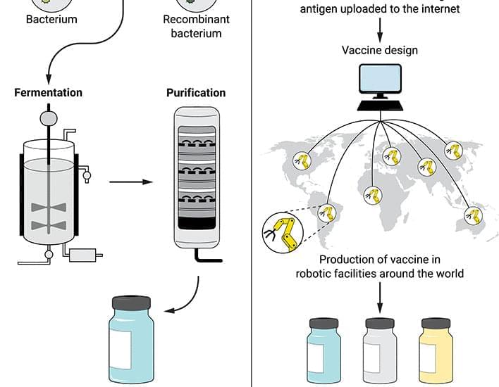
The SARS-CoV-2 pandemic has generated a renaissance in vaccinology, with COVID-19 mRNA vaccines delivering a “digital code” of the viral antigen with no need to purify proteins or inactivate pathogens.
One of the reasons branching identity is being accepted more seriously these days.
Beginning in the 1950s, experiments with split-brain patients revealed that consciousness could be divided between the two hemispheres of the brain. A surprising implication was that if consciousness could be divided, then it could also be combined. Evidence of this came in 2006, when conjoined twins Krista and Tatiana Hogan were born. The Hogan sisters were born with their brains connected by a thalamic bridge, which allowed a unique mental connection between them. We explore this surprising mental connection, and the possibility that we may one day connect our own minds with other conscious beings, together with what this might mean for our concept of self, identity, and the future of mind.
Please consider supporting my work on Patreon here ~ https://www.patreon.com/wakingcosmos.
Thank you!
This documentary is a non-profit, educational film. All footage used with permission, or in compliance with fair use/fair dealing.
Music by Scott Buckley and Aleks Michalski.
One promising solution to plastic pollution is mycelium or mushroom packaging. It is made of 2 ingredients: mushrooms and hemp. Mycelium is the underground network of very durable, thread-like filaments called hyphae. It is mixed with agricultural waste like wood chips, oat hulls, cotton burrs or hemp hurds.
Link to my Patreon page: https://www.patreon.com/Belinda_Carr.
Chapters.
0:00 Introduction.
1:00 How its made.
2:43 Products.
4:32 Advantages.
5:30 Disadvantages.
6:19 Myths.
7:12 Conclusion.
One of the largest mushroom packaging manufacturers in the world is Ecovative Design, a New York based biotech company founded in 2006. They sent me these samples of their product. Their manufacturing process is pretty straight forward.
Their designers create a 3D CAD model of custom packaging.
A CNC machine routes the design into MDF.
Plastic trays are thermoformed around the MDF pieces.
The tray is filled with their proprietary hemp hurd and mycelium blend.
It is allowed to grow for 4 days in a controlled environment with regulated temperatures, airflow, CO2 and humidity levels.
It is popped out of tray and allowed to continue growing for 2 more days to create a velvety layer of overgrowth.
The packaging is then heat treated to dry out, kill spores and stop the growth process.
This material can last for 30 years in dry, temperature controlled indoor environments. It is also 100% biodegradable and a nutrient for soils and plants. When broken down into 1 cubic centimeter pieces, it will compost in just 45 days. In the ocean, it will compost in 180 days.

Roche and its Genentech subsidiary have committed up to $12 billion to Recursion in return for using its Recursion Operating System (OS) to advance therapies in 40 programs that include “key areas” of neuroscience and an undisclosed oncology indication.
Recursion OS applies machine learning and high-content screening methods in what the companies said would be a “transformational” model for tech-enabled target and drug discovery.
The integrated, multi-faceted OS is designed to generate, analyze and glean insights from large-scale proprietary biological and chemical datasets—in this case, extensive single-cell perturbation screening data from Roche and Genentech—by integrating wet-lab and dry-lab biology at scale to phenomically capture chemical and genetic alterations in neuroscience-related cell types and select cancer cell lines.
Monash University, Australia scientists have discovered an enzyme that is key to why exercise improves our health. Importantly this discovery has opened up the possibility of drugs to promote this enzyme’s activity, protecting against the consequences of aging on metabolic health, including type 2 diabetes.
The proportion of people worldwide over 60 years old will double in the next three decades and by 2031, more than six million Australians will be over 65 years old. The incidence of type 2 diabetes increases with age so this aging population will also result in an increased incidence of the disease globally.
One of the main reasons for the increased prevalence of type 2 diabetes with age is the development of insulin resistance, or an inability for the body to respond to insulin, and this is often caused by reduced physical activity as we age.
ROCHESTER, Minn. ― Adding messenger RNA, or mRNA therapy improves the response to cancer immunotherapy in patients who weren’t responding to the treatment, Mayo Clinic research shows. Immunotherapy uses the body’s immune system to prevent, control and eliminate cancer. The study is published in Cancer Immunology Research, a journal of the American Association for Cancer Research.
The phrase messenger RNA and its acronym, mRNA, have become familiar to the public during the COVID-19 pandemic. The mRNA vaccines for COVID-19 work by instructing cells in the body how to make a protein that triggers an immune response against the virus.
MRNA technology has also been of interest to cancer researchers and physicians. One of the major obstacles in cancer treatment is the low response rate in patients who receive immune checkpoint inhibitors to prevent an immune response from being so strong that it destroys healthy cells in the body.
In this video Dr. Lustgarten introduces his N of 1 experiment and gives an overview of the processes that he follows. He also discusses why he thinks it is important to track your own markers and not just rely on published trials.
Dr. Michael Lustgarten is a scientist at the Tufts University Human Nutrition Research Center on Aging in Boston, Massachusetts. His research currently focuses on the role of the gut microbiome and serum metabolome on muscle mass and function in older adults.
In this series of interviews Dr Lustgarten shares his experience with his rigorous n of 1 experiment over the last 7 years and shows how we or anyone can conduct a similar trial by tracking food, exercise and sleep, measure results and derive relationships between them, with a goal of extending our healthspan.
Dr Lustgarten’s channel on YouTube: https://www.youtube.com/channel/UCT1UMLpZ_CrQ_8I431K0b-g.
***************************************************************
Health claims Disclosure: Information provided on this video is not a substitute for direct, individual medical treatment or advice. Please consult with your doctor first. Products or services mentioned in this video are not a recommendation.
Audio Copyright Disclaimer.
Please note that we have full authorization to the music that we used in our videos as they were created using the service WeVideo which provides the rights to the music. The rights are detailed in the terms of use that can be reviewed here https://www.wevideo.com/terms-of-use and any following inquiries should be addressed to [email protected].
*****************************************************************

Abstract. The cnidarian model organism Hydra has long been studied for its remarkable ability to regenerate its head, which is controlled by a head organizer located near the hypostome. The canonical Wnt pathway plays a central role in head organizer function during regeneration and during bud formation, which is the asexual mode of reproduction in Hydra. However, it is unclear how shared the developmental programs of head organizer genesis are in budding and regeneration. Time-series analysis of gene expression changes during head regeneration and budding revealed a set of 298 differentially expressed genes during the 48-h head regeneration and 72-h budding time courses. In order to understand the regulatory elements controlling Hydra head regeneration, we first identified 27,137 open-chromatin elements that are open in one or more sections of the organism body or regenerating tissue. We used histone modification ChIP-seq to identify 9,998 candidate proximal promoter and 3,018 candidate enhancer-like regions respectively. We show that a subset of these regulatory elements is dynamically remodeled during head regeneration and identify a set of transcription factor motifs that are enriched in the enhancer regions activated during head regeneration. Our results show that Hydra displays complex gene regulatory structures of developmentally dynamic enhancers, which suggests that the evolution of complex developmental enhancers predates the split of cnidarians and bilaterians.
