Toggle light / dark theme

Turmeric and ginger extract may boost implant bonding and kill 92% bacteria

An extract of turmeric and ginger helps bone implants bond strongly while killing bacteria and cancer cells, according to new research from Washington State University with implications for millions of patients with joint replacements and bone cancer. In early tests, the extract roughly doubled bone bonding within six weeks around the implant site, killed more than 90% of bacteria on implant surfaces, and sharply reduced cancer-causing cells. The findings marry elements of a naturopathic approach drawing on traditional medicine with current medical technologies. Turmeric, a golden-orange spice, and ginger root have been used for food and medicinal purposes in China and India for thousands of years.

“Basically, I say it’s combining the best with the latest,” said Susmita Bose, the Westinghouse Distinguished Chair Professor in WSU’s School of Mechanical and Materials Engineering and corresponding author of the paper. “The best part is from the food, and the latest aspect comes from the biomedical device.”

The new study, published in the Journal of the American Ceramic Society, is the most recent work from Bose and Amit Bandyopadhyay, Boeing Distinguished Professor in the School of Mechanical and Materials Engineering, demonstrating that compounds from turmeric and ginger can be effective supplements to cutting-edge medical treatment. That work builds upon their earlier research into the use of 3D printing to produce bone implants, an idea once considered far-fetched that is now a common way to manufacture implants.

3D microscopy reveals how a tick-borne virus reshapes human cells to replicate

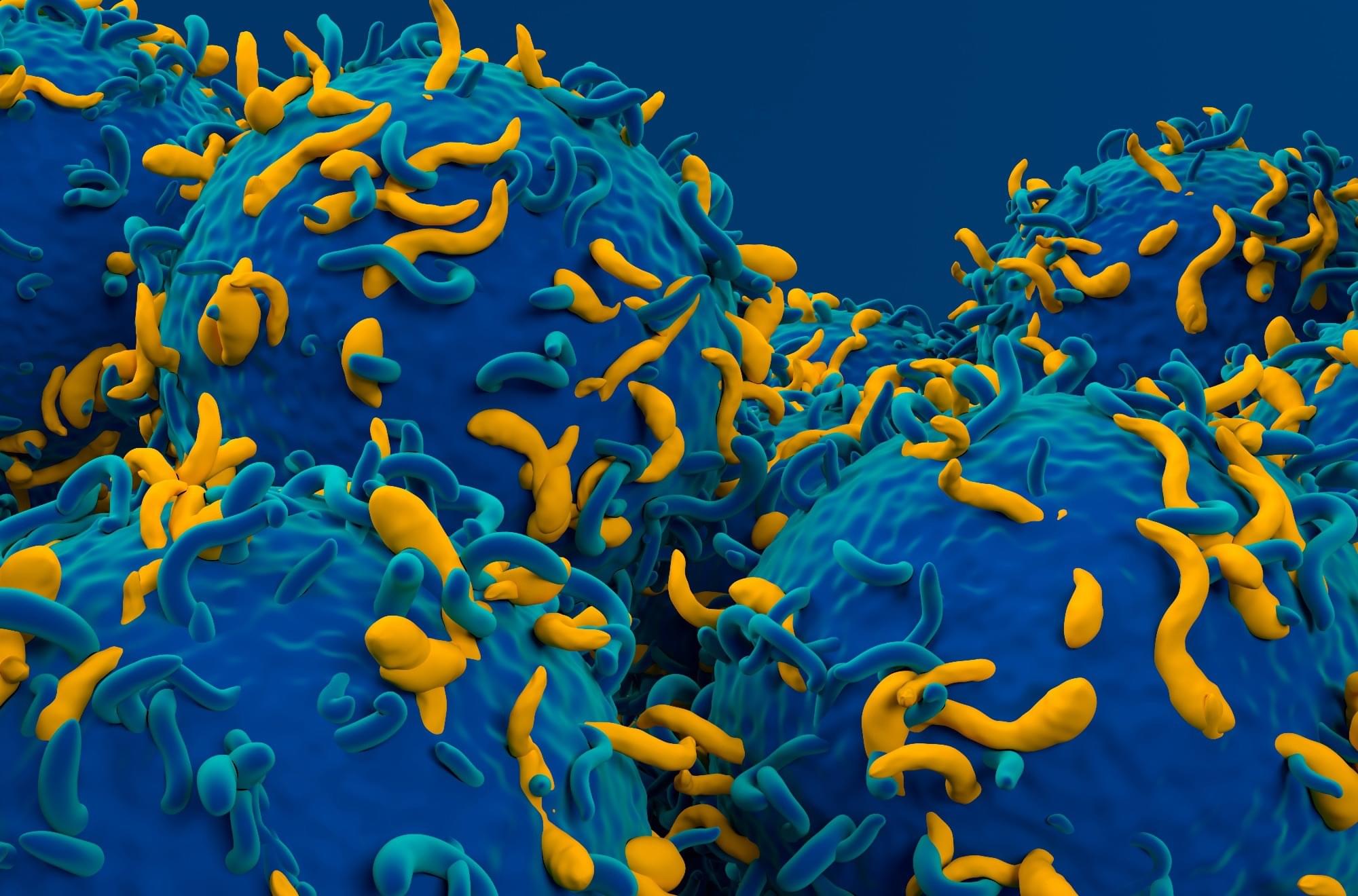
Researchers at Umeå University show how tick-borne viruses remodel human cells into virus factories, using an advanced microscopy method. The findings provide new insight into how the virus replicates and matures, knowledge that may become important for future treatments against TBE. The study is published in Nature Communications.

“When we saw the three-dimensional images for the first time, we immediately realized how much new information we could gain about the virus’s replication,” says Lars-Anders Carlson, professor at the Department of Medical Chemistry and Biophysics at Umeå University, who led the study.

One of the most dangerous viral diseases spread in Europe is tick-borne encephalitis. A bite from an infected tick can transmit the TBE virus to humans and cause severe inflammation of the brain. Using electron microscopy, researchers at Umeå University have now discovered how tick-borne viruses reshape infected human cells and turn them into virus factories.

What this AI epitope library means for vaccines, immunotherapy and biosensors

A new tool makes it possible to screen millions of tiny protein fragments and select those that can be recognized by the immune system. The CIC biomaGUNE Center for Cooperative Research in Biomaterials has developed epiGPTope, a system that uses machine learning to generate and classify epitopes, in collaboration with the company Multiverse Computing.

The immune system is triggered by the presence of viruses or bacteria. When the antibodies produced recognize the epitopes, a small part of these viruses or bacteria, they launch an attack strategy. These epitopes are small fragments of protein recognized by antibodies or by immune cell receptors. So discovering new epitope sequences that target specific antibodies is essential for the development of diagnostic tools, immunotherapies and vaccines.

CIC biomaGUNE’s Biomolecular Nanotechnology laboratory, led by the Ikerbasque Research Professor Aitziber L. Cortajarena, is creating a library or database of hundreds of thousands of synthetic epitopes using this AI-based technique. The work is published in the journal ACS Synthetic Biology.

A layered approach sharpens brain signals in optical imaging

Near-infrared spectroscopy, or fNIRS, offers a way to monitor brain activity without surgery or radiation by tracking changes in blood flow and oxygenation. Light sources placed on the scalp send near-infrared light into the head, and detectors measure the light that scatters back. Because this light must pass through the scalp and skull before reaching the brain, the measured signal always includes a mix of superficial and cerebral contributions. Separating those signals has long been a central challenge for fNIRS researchers.

In a study published in Biophotonics Discovery, researchers from the Tufts University Diffuse Optical Imaging of Tissue Laboratory show that combining a specific source–detector geometry with a simple, anatomically informed tissue model can substantially improve how fNIRS data are interpreted.

By accounting for how light travels through layered head structures, the approach makes it possible to better isolate brain-specific signals without relying on complex imaging systems or subject-specific MRI scans.

Tiny Robots Built From DNA Could Hunt Viruses and Deliver Drugs Inside the Human Body

For most of human history, medical treatment has relied on methods such as pills, injections, and surgery. Now, scientists are exploring a new idea: making tiny, programmable machines from DNA that can move through the bloodstream.

A recent review published in the journal SmartBot says these DNA nanorobots could one day be capable of delivering drugs to specific locations in the body, capturing viruses like SARS-CoV-2, and even helping build tiny computing devices. Even though these ideas are exciting, the technology is still in its early stages.

Early Stages of Development.

What keeps vision cells alive?

Clear patterns emerged: two kinase inhibitors consistently protected cones over extended periods.

The researchers identified inhibitors of casein kinase 1 (CK1) that protected cones, heat shock protein 90 (HSP90) inhibitors that saved cones in the short term but damaged them in the longer term, and broad histone deacetylase (HDAC) inhibition by many compounds that significantly damaged cones.

The protective effects held across different stress conditions and were further confirmed in a mouse model of retinal degeneration, supporting their broader relevance.

Beyond identifying protective pathways, the study makes a comprehensive dataset publicly available, covering the compounds tested, their molecular targets, and their effects on human cone survival. This resource will guide the development of therapies aimed at preserving central vision and enable a systematic assessment of potential retinal toxicity. ScienceMission sciencenewshighlights.


Scientists have identified genetic pathways and compounds capable of protecting cone photoreceptors from the degeneration that underlies conditions like age-related macular degeneration.

Cone photoreceptors, concentrated in the macula, are essential for reading, recognizing faces, and perceiving colors. Their death, as it happens in many inherited retinal diseases and macular degeneration, leads to the loss of central vision. Despite decades of research, no approved therapies can halt this process. This new study, conducted by researchers addresses this unmet need using a human-based experimental system.

CBD found to reverse brain damage in mouse model of Alzheimer’s disease

Alzheimer’s disease (AD) is a neurodegenerative disorder characterized by progressive memory loss, cognitive decline, and behavioral changes. The deficits linked to AD are known to result from the abnormal accumulation of proteins, particularly tau and β-amyloid (Aβ) in the brain and between nerve cells, which causes neuroinflammation and can prompt the degradation of brain cells.

The non-psychoactive compound derived from the Cannabis sativa plant, called cannabidiol (CBD), was recently found to show promise for protecting brain cells from damage.

Compared to Δ9-tetrahydrocannabinol (THC), the compound in cannabis that elicits feelings of euphoria and alters a user’s mental state, CBD is safer and could thus be easier to introduce in clinical settings.

Scientists discover hidden brain switch that tells you to stop eating

Your brain’s “stop eating” signal may come from an unexpected source. Researchers found that astrocytes—once thought to just support neurons—actually play a key role in controlling appetite. After a meal, glucose triggers tanycytes, which send signals to astrocytes that then activate fullness neurons. This newly discovered pathway could lead to innovative treatments for obesity and eating disorders.

/* */