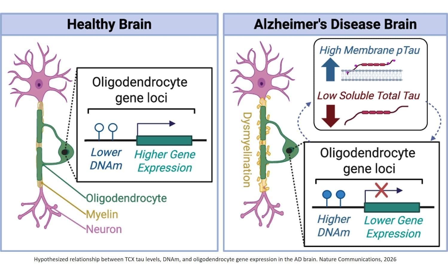
Toggle light / dark theme

Scientists from the Biology Centre of the Czech Academy of Sciences found forty new freshwater viruses infecting aquatic microorganisms this year. The first one, which they isolated and described in detail, was named Budvirus after the South Bohemian capital České Budějovice. It belongs to “Giant Viruses” and it infects unicellular algae called cryptophytes.

Researchers have confirmed that this virus has an important role in the ecosystem, as it controls algal bloom, helping to maintain balance in the aquatic environment. The discoveries of all the viruses were made at the Římov reservoir near České Budějovice, which has been regularly monitored by South Bohemian hydrobiologists for five decades and is one of the most studied freshwater reservoirs in Europe. The work is published in The ISME Journal.

Although we have freshwater ecosystems such as lakes, ponds, reservoirs and rivers all around us, their microscopic representatives, especially viruses and bacteria, are still a little-explored area. A drop of water can contain a million bacteria and ten times more viruses, but only a handful of them have been described. Recent methods, such as environmental DNA analysis, are making great strides in the study of the aquatic microworld. This is also one of the methods used by the Czech scientific team.

Our bodies divest themselves of 60 billion cells every day through a natural process of cell culling and turnover called apoptosis.

These cells — mainly blood and gut cells — are all replaced with new ones, but the way our bodies rid themselves of material could have profound implications for cancer therapies in a new approach developed by Stanford Medicine researchers.

They aim to use this natural method of cell death to trick cancer cells into disposing of themselves. Their method accomplishes this by artificially bringing together two proteins in such a way that the new compound switches on a set of cell death genes, ultimately driving tumor cells to turn on themselves. The researchers describe their latest such compound in a paper published Oct. 4 in Science.

Sometimes there are slightly different versions, or sequences of genes. There are several versions of the apolipoprotein E (APOE) gene, for example. One of them, called APOE4, has been linked to a much higher risk of developing Alzheimer’s disease, and carriers often have worse forms of the disease compared to carriers of other forms like APOE3. There are immune cells in the brain called microglia that help protect the brain from damage and harm. But when APOE4 is expressed, microglia seem to start to cause inflammation, and misfolded proteins to form in the brain, which can lead to serious problems. The findings have been reported in Cell Stem Cell.

In this work, the researchers developed a mouse model that could generate the human APOE4 protein in their brains. Next, the investigators eliminated microglia from these mouse brains. The formation of two misfolded proteins that are hallmarks of Alzheimer’s diseases: amyloid and tau, was halted.

There are many processes and proteins that help the body fight a flu infection. One of them is known as IFITM3. Researchers have now shown that this protein can help prevent viruses from mutating after they have infected a new host. But some people are deficient in IFITM3, which can raise their risk of a severe flu infection. That deficiency is not unusual in some groups. For example, around twenty percent of Chinese people and four percent of people with European ancestry carry variants in IFITM3 that can interfere with the protein’s expression. This study has shown that these genetic variants can allow flu viruses to establish infections even when the virus is present at very low levels that would not usually cause infection. The findings have been reported in Nature Communications.

The IFITM3 (interferon-induced transmembrane protein 3) protein is part of the innate immune system, and is generated at high levels after the detection of a flu infection. It can sequester viral particles so that they are not able to replicate, which reduces the severity of flu infections. Mouse models that are IFITM3 deficient are extremely vulnerable to the flu.

Summary: AI models trained on MRI data can now distinguish brain tumors from healthy tissue with high accuracy, nearing human performance. Using convolutional neural networks and transfer learning from tasks like camouflage detection, researchers improved the models’ ability to recognize tumors.

This study emphasizes explainability, enabling AI to highlight the areas it identifies as cancerous, fostering trust among radiologists and patients. While slightly less accurate than human detection, this method demonstrates promise for AI as a transparent tool in clinical radiology.

A big day of output for the Human Cell Atlas, a global collaborative project with 100 countries to understand our ~37 trillion cells A Wikipedia of our cells, a “remarkable achievement” https://nature.com/articles/d41586-024-03754-y


In a collection of research articles and related content, the Human Cell Atlas consortium presents tools, data and ideas towards the generation of their first draft atlas of cells in the human body.

Join us on Patreon! https://www.patreon.com/MichaelLustgartenPhD

Discount Links/Affiliates:
Blood testing (where I get my labs): https://www.ultalabtests.com/partners/michaellustgarten.

Clearly Filtered Water Filter: https://get.aspr.app/SHoPY

At-Home Metabolomics: https://www.iollo.com?ref=michael-lustgarten.
Use Code: CONQUERAGING At Checkout.

Epigenetic, Telomere Testing: https://trudiagnostic.com/?irclickid=U-s3Ii2r7xyIU-LSYLyQdQ6…M0&irgwc=1
Use Code: CONQUERAGING

NAD+ Quantification: https://www.jinfiniti.com/intracellular-nad-test/

Genes aren’t the sole driver instructing cells to build multicellular structures, tissues, and organs. In a paper published in Nature Communications, USC Stem Cell scientist Leonardo Morsut and Caltech computational biologist Matt Thomson characterize the influence of another important developmental driver: cell density, or how loosely or tightly cells are packed into a given space.

In both computational models and laboratory experiments, the team of scientists used cell density as an effective tool for controlling how pattern themselves into complex structures.

“This paper represents progress towards our big picture goal of engineering synthetic tissues,” said Morsut, an assistant professor of stem cell biology and regenerative medicine, and biomedical engineering at the Keck School of Medicine of USC.

Summary: The Human Cell Atlas (HCA) consortium has published over 40 studies revealing groundbreaking insights into human biology through large-scale mapping of cells. These studies cover diverse areas such as brain development, gut inflammation, and COVID-19 lung responses, while also showcasing the power of AI in understanding cellular mechanisms.

By profiling over 100 million cells from 10,000 individuals, HCA is building a “Google Maps” for cell biology to transform diagnostics, drug discovery, and regenerative medicine. The initiative emphasizes diversity, including underrepresented populations, to ensure a globally inclusive understanding of health and disease.

Summary: Autism-linked SHANK3 gene mutations disrupt not only neurons but also oligodendrocytes, essential for producing myelin, which insulates nerve fibers. This damage reduces brain signal efficiency and impairs behavior.

Using gene therapy, researchers successfully repaired these cells in a mouse model, restoring their function and myelin production. They validated their findings with human-derived stem cells, confirming similar impairments and repair mechanisms.

This discovery highlights a significant role for oligodendrocytes in autism and opens the door for innovative treatments targeting myelin dysfunction. The study underscores both the biological complexity of autism and the promise of genetic therapies for intervention.