Converting DNA sequences and particle vibrations into notes allows researchers to recognize unseen patterns and create songs for outreach.
Category: biotech/medical – Page 2,048
UMN research shows people can control robotic arm with their minds
Researchers at the University of Minnesota have made a major breakthrough that allows people to control a robotic arm using only their minds. The research has the potential to help millions of people who are paralyzed or have neurodegenerative diseases.
The study is published online today in Scientific Reports, a Nature research journal.
To read the full research paper, entitled “Noninvasive Electroencephalogram Based Control of a Robotic Arm for Reach and Grasp Tasks,” visit the Nature Scientific Reports website.
Groundbreaking study demonstrates potential to help millions of people with disabilities.
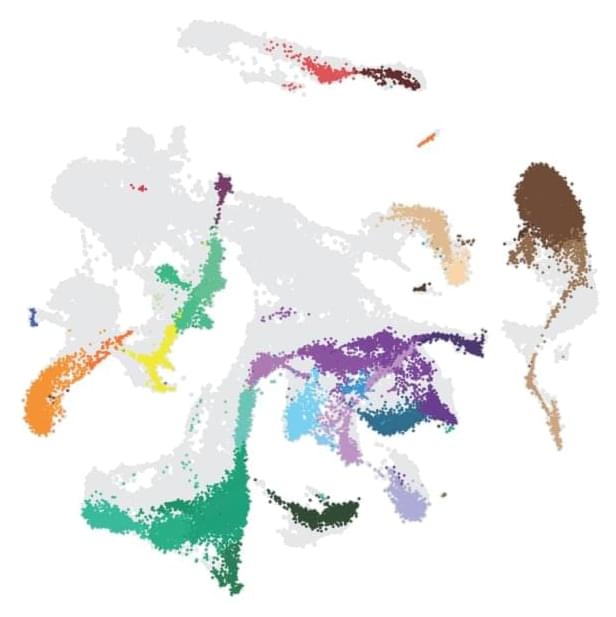
Single Cell Analysis Technologies Help Generate Unprecedented Maps of Disease
Astronomy was born when early scientists peered into the sky with their naked eyes and recorded what they could see above them. Then, the invention of the telescope brought forth new insights. And today, astronomers conduct their studies from big observatories and launch sophisticated telescopes into space for a much more in-depth look.
Now, a similar evolution is occurring in biology as scientists develop new techniques for taking a closer look at cells—the basic living units of organs. The origins of cell biology date back to 1,665 when Robert Hooke was the first to look at a cell under a simple compound microscope. But while the development of more powerful microscopes such as the scanning electron microscope has allowed scientists to take a peek at molecules smaller than a billionth of a meter, until recently they have never had the ability to look at the molecular profile of a single cell.
Yale researchers across disciplines are using single cell technologies to profile various kinds of cells that exist together in both healthy and diseased organs and create the most detailed blueprints of diseases to date, as well as to better understand how various cells develop over time and interact with one another. Through creating these “cell atlases” of organs throughout the body, they hope to shed light on the mechanisms of a wide variety of diseases and biological development.

Scientists develop real-life mind controllable robotic hand using AI
This AI powered prosthetic arm understands what you think. Muscle-controlled prosthetic limbs that patients with amputations across the globe currently use have various limitations and challenges. Good quality prosthetics parts are cumbersome, come with a complex setup, and require patients to undergo training for several months to learn their use. Interestingly, a new technology proposed by a team of researchers at the University of Minnesota (UMN) can overcome all such challenges.
It may sound like science-fiction, but the researchers claim that the new technology would allow patients to control robotic body parts using their thoughts. By employing artificial intelligence and machine learning, the researchers at UMN have developed a portable neuroprosthetic hand. The robotic hand comes equipped with a nerve implant linked to the peripheral nerve in a patient’s arm.
Explaining the significance of their neuroprosthetic innovation, project collaborator and UMN neuroscientist Edward Keefer said, “We are well along the way toward allowing upper limb amputees at least, and other people in the future, to have totally natural and intuitive control of their prosthetic devices.” ## THE NEUROPROSTHETIC HAND IS DIFFERENT FROM YOUR REGULAR PROSTHETIC LIMBS
The prosthetic body parts currently available on the market detect shoulder, chest, or muscle movement. They have sensors to recognize signals in specific regions of the human body. Therefore, every time a patient wants to move his hand, he is required to trigger his body muscles. Adapting to such muscle-driven limb movement is not easy for patients, and many such devices are not suitable for physically weak individuals.
Some advanced and efficient muscle-sensitive prosthetics come with complex wiring and other arrangements that make them difficult to use. The amputees have to go through a lot of training to adjust to such devices, which often increases frustration and stress. Now imagine a device that starts working immediately, is less invasive, requires no training, no muscle activation, and no complex setup.
The neuroprosthetic arm enables the patients to move their arms simply at the will of their minds. It is an efficient, easy to use, and a lot more intuitive alternative to any commercial prosthetic system available.
Dr. Maria Van Kerkhove, PhD — Emerging Diseases and Zoonoses Unit Head — World Health Organization
Preparedness For Emerging Diseases & Zoonoses — Dr. Maria Van Kerkhove, Ph.D., Emerging Diseases and Zoonoses Unit Head, World Health Organization, (WHO)
Dr. Maria Van Kerkhove, Ph.D., (https://www.imperial.ac.uk/people/m.vankerkhove) is an infectious disease epidemiologist who serves as the technical lead for the COVID-19 response at the World Health Organization (https://www.who.int/en/), where she develops guidance, training programs, and information products for the continuously evolving state of the pandemic, as well serving as the Emerging Diseases and Zoonoses Unit Head.
Dr. Van Kerkhove began her journey in global health given her interest in viruses and how they infect and impact both humans and animals. She received her undergraduate degree in biological sciences from Cornell University, her master’s degree in epidemiology from Stanford University, and a PhD in infectious disease epidemiology from the London School of Tropical Hygiene and Medicine where she authored her PhD on pathogenic avian influenza H5N1 in Cambodia.
Following her PhD, Dr. Van Kerkhove was a postdoctoral researcher with the WHO and acted as a liaison for the Imperial College London’s Medical Research Council Centre for Outbreak Analysis.
Dr. Van Kerkhove continued working with the WHO and prior to COVID-19, was serving as the MERS-CoV Technical Lead in addition to being the Unit Head for the Emerging Disease and Zoonoses Unit. Her focus in these areas includes developing prevention and control programs around high threat respiratory pathogens.
Dr. George Church—Gene Therapy and Aging
In this episode of Longevity by Design, our hosts, Dr. Gil Blander and Ashley Reaver, MS, RD, CSSD, are joined by Dr. George Church, Professor of Genetics at Harvard Medical School. Tune in as Dr. George Church discusses the many roles of gene therapy, including its ability to reverse age-related diseases.
For science-backed ways to live a healthier, longer life, download InsideTracker’s InnerAge eBook at insidetracker.com/podcast.
Social:
Instagram — https://www.instagram.com/insidetracker.
Twitter — https://twitter.com/InsideTracker.
Facebook — https://www.facebook.com/InsideTracker.
Website — https://www.insidetracker.com/
In the Press — https://www.insidetracker.com/press-page/
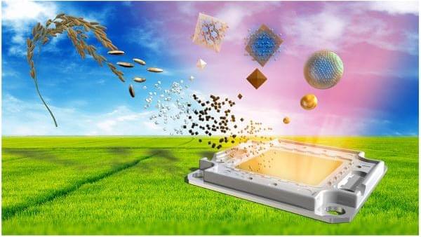
World-first quantum dot LED lights made from discarded rice husks
From TVs, to solar cells, to cutting-edge cancer treatments, quantum dots are beginning to exhibit their unique potential in many fields, but manufacturing them at scale would raise some issues concerning the environment. Scientists at Japan’s Hiroshima University have demonstrated a greener path forward in this area, by using discarded rice husks to produce the world’s first silicon quantum dot LED light.
“Since typical quantum dots often involve toxic material, such as cadmium, lead, or other heavy metals, environmental concerns have been frequently deliberated when using nanomaterials,” said Ken-ichi Saitow, lead study author and a professor of chemistry at Hiroshima University. “Our proposed process and fabrication method for quantum dots minimizes these concerns.”
The type of quantum dots pursued by Saitow and his team are silicon quantum dots, which eschew heavy metals and offer some other benefits, too. Their stability and higher operating temperatures makes them one of the leading candidates for use in quantum computing, while their non-toxic nature also makes them suitable for use in medical applications.
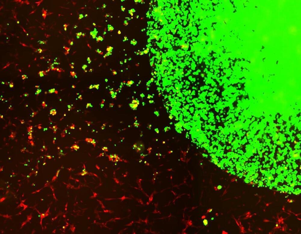
“Off the Shelf” Engineered Stem Cells Created To Treat Aggressive Brain Cancer
Investigators uncovered a diagnostic method to identify receptors on cancer cells in the blood, then engineered a cell-based therapy to target and kill tumor cells in the brain, paving the way to clinical testing.
Glioblastomas (GBMs) are highly aggressive cancerous tumors of the brain and spinal cord. Brain cancers like GBM are challenging to treat because many cancer therapeutics cannot pass through the blood-brain barrier, and more than 90% of GBM tumors return after being surgically removed, despite surgery and subsequent chemo-and radiation therapy being the most successful way to treat the disease. In a new study led by investigators at Brigham and Women’s Hospital and Harvard Medical School, scientists devised a novel therapeutic strategy for treating GBMs post-surgery by using stem cells taken from healthy donors engineered to attack GBM-specific tumor cells. This strategy demonstrated profound efficacy in preclinical models of GBM, with 100 percent of mice living over 90 days after treatment. Results will be published today (May 19, 2022) in the journal Nature Communications.
“This is the first study to our knowledge that identifies target receptors on tumor cells prior to initiating therapy, and using biodegradable, gel-encapsulated, ‘off-the-shelf’ engineered stem cell based therapy after GBM tumor surgery,” said Khalid Shah, MS, PhD, director of the Center for Stem Cell and Translational Immunotherapy (CSTI) and the vice chair of research in the Department of Neurosurgery at the Brigham and faculty at Harvard Medical School and Harvard Stem Cell Institute (HSCI).

1st monkeypox case in US this year reported in Massachusetts
A Massachusetts resident has tested positive for monkeypox, health officials confirmed Wednesday, making it the first case of the rare virus detected in the United States this year.
According to a release from the Massachusetts Department of Public Health, the patient is an adult male who recently traveled to Canada. The department completed initial testing Tuesday and was confirmed by the Centers for Disease Control and Prevention.
“The case poses no risk to the public, and the individual is hospitalized and in good condition,” MDPH stated in a press release. “DPH is working closely with the CDC, relevant local boards of health, and the patient’s health care providers to identify individuals who may have been in contact with the patient while he was infectious.”