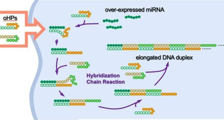
This was the first time scientists were able to develop a hairpin-shaped DNA strand that can activate a natural immune response to target and kill specific cancerous cells. Tuesday 27 December 2022 10:48 A new way of using DNA to kill cancer cells which could pave the way for a cure for the disease has been created by scientists.
In November 2016, virologist David Evans traveled to Geneva for a meeting of a World Health Organization committee on smallpox research. The deadly virus had been declared eradicated 36 years earlier; the only known live samples of smallpox were in the custody of the United States and Russian governments.
Evans, though, had a striking announcement: Months before the meeting, he and a colleague had created a close relative of smallpox virus, effectively from scratch, at their laboratory in Canada. In a subsequent report, the WHO wrote that the team’s method “did not require exceptional biochemical knowledge or skills, significant funds, or significant time.”
Evans disagrees with that characterization: The process “takes a tremendous amount of technical skill,” he told Undark. But certain technologies did make the experiment easier. In particular, Evans and his colleague were able to simply order long stretches of the virus’s DNA in the mail, from GeneArt, a subsidiary of Thermo Fisher Scientific.
Five of our favourite interviews with thought leaders and investors on the opportunity presented by longevity.
From spending billions on research to calls for fundamental changes to way we deliver healthcare, this year we heard from a host of thought leaders who shared their views on how to make longevity a reality. Today we bring you five of the best.
When we spoke to Professor Sir John Bell, we expected to learn more about a new UK initiative to study of the health of five million citizens to enable more effective ways to prevent, detect and treat diseases. But what we got was a stirring call to action for a change in the way healthcare is conducted.
When the COVID-19 pandemic struck and work was forced to go remote, services like Zoom and Microsoft Teams rose to the occasion to keep people connected and work ongoing. For many, work turned into a series of online meetings to be attended day after day.
Blood pressure genetic risk score can predict risk of heart attacks and stroke.
Nearly half of all American adults have elevated blood pressure or hypertension and high blood pressure contributes to 65 percent of cardiovascular deaths in the US. Now researchers at University of Alabama at Birmingham have used genomic information to create a blood pressure “genetic risk score”.
Longevity. Technology: Cardiovascular disease is the leading cause of death worldwide and is responsible for a significant burden of morbidity and mortality. As people age, their risk of developing CVD increases, making it a major contributor to the morbidity and mortality associated with aging. That there is a pressing need for research into CVD in order to identify effective strategies for prevention and treatment would seem obvious, but this research is particularly important as the global population is aging and the prevalence of CVD is expected to rise with it. However, having such an enormous number of people at risk brings extra problems – how can risks be quantified and determined on an individual basis? The answer could lie in understanding and leveraging genetic data.
Professor Carmit Levy. Credit: Tel Aviv University.
Professor Carmit Levy from the Department of Human Genetics and Biochemistry and Dr. Yftach Gepner from the School of Public Health and the Sylvan Adams Sports Institute at TAU’s Sackler Faculty of Medicine conducted the study. Prof. Levy notes that the new research has resulted in a very important discovery by merging scientific know-how from different schools at TAU, which may help avoid metastatic cancer, Israel’s top cause of death. The study was recently published on the cover of the journal of Cancer Research.
Prof. Levy and Dr. Gepner: “Studies have demonstrated that physical exercise reduces the risk for some types of cancer by up to 35%. This positive effect is similar to the impact of exercise on other conditions, such as heart disease and diabetes. In this study we added new insight, showing that high-intensity aerobic exercise, which derives its energy from sugar, can reduce the risk of metastatic cancer by as much as 72%. If so far the general message to the public has been ‘be active, be healthy’, now we can explain how aerobic activity can maximize the prevention of the most aggressive and metastatic types of cancer.”
Almost every patient who gets myeloma and is treated with a standard therapy also experiences relapse. But researchers have developed an antibody therapy that triggers the immune system to destroy these cancer cells. The bispecific antibody can bind to T cells and multiple myeloma cells at once, to kill the cancer. This immunotherapy, called talquetamab, was astonishingly effective, and worked in about 73 percent of patients who were treated with the drug in two clinical trials. The treatment even helped for a patient who had a cancer that resisted all therapies that have been approved for multiple myeloma.
Talquetamab takes advantage of a receptor on myeloma cells called GPRC5D, and CD3, a complex and co-receptor on the surface of T cells. Anti-CD3 antibodies have long been known to cause the activation of T cells. Mouse studies showed that talquetamab can recruit and activate CD3-positive T-cells, which inhibits the formation and growth of tumors.
The Food and Drug Administration (FDA) considers aging to be a natural process. This makes it difficult to get FDA approval for drugs that seek to slow or reverse the biological process of aging. Instead, drugs intended to target aging must target a disease that often results from the aging process in order to demonstrate efficacy and gain approval.
But there is growing consensus and effort among scientists to convince the FDA that aging itself should be classified as a disease and an appropriate target for drug development.