A team of scientists from the University of Pittsburgh School of Medicine has found the missing puzzle piece in the mystery of how melanoma tumors control their mortality.
In a paper published in Science, they describe how they identified the specific genetic changes that allow tumors to grow rapidly while also preventing their own death. This discovery could have significant implications for the way melanoma is understood and treated by oncologists.
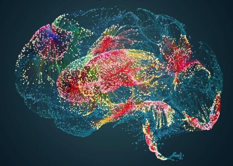
Summary: Roflumilast, a drug commonly prescribed for the treatment of COPD and asthma helps retrieve learning memories following a period of sleep deprivation in mice.
Source: University of Groningen.
Students sometimes pull an all-nighter to prepare for an exam. However, research has shown that sleep deprivation is bad for your memory.
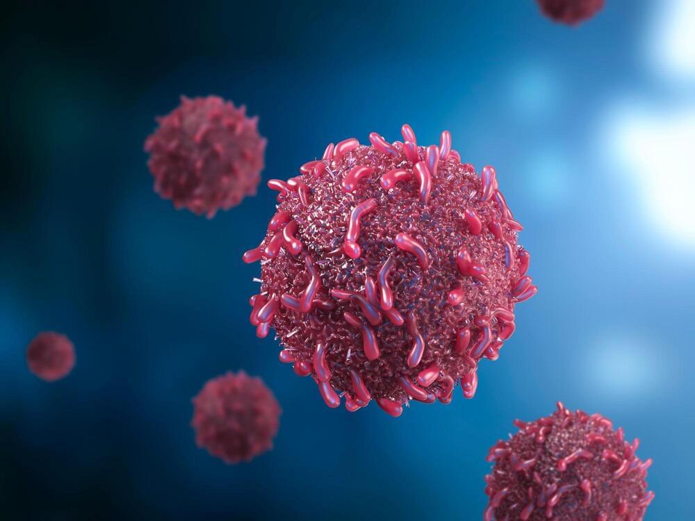
A protein that helps lethal skin cancer spread through the body has been identified, according to scientists, offering new hope for cancer treatment. Protein LAP1 allows cancer cells to become more aggressive by letting them change the shape of their nucleus and migrate around the body. The most serious type of cancer cells, melanoma, was found to harbour LAP1 and high levels of it were linked to poor prognosis.
Pioneering Hematopoietic Stem Cell Transplants (HSCT) For Autoimmune Disorders — Dr. Richard K. Burt
Dr. Richard K. Burt MD (https://astemcelljourney.com/about/drrichardburt/) is a Fulbright Scholar, Professor of Medicine at Scripps Health Care, tenured retired Professor of Medicine at Northwestern University where he served as Chief of Immunotherapy and Autoimmune Diseases, and CEO of Genani Biotechnology.
Dr. Burt endeavored for thirty-five years, first with animal models and then with some of the world’s first clinical trials, to bring the field of stem cell and cellular therapy to the patient’s bedside.
Dr. Burt has published more than 145 mostly first author articles and is the Editor of four medical textbooks. He was the first Autoimmune Committee Chairperson for the International Bone Marrow Transplant Registry (IBMTR) and was the principal investigator of a National Institute of Health (NIH) $10,000,000 multi-center contract to develop stem cell clinical trials for autoimmune diseases.
Dr. Burt performed America’s first hematopoietic stem cell transplant (HSCT) for multiple sclerosis (MS), systemic lupus erythematosus (SLE), Crohn’s disease (CD), stiff person syndrome (SPS), and chronic inflammatory demyelinating polyneuropathy (CIDP) and published the world’s first randomized clinical stem cell transplantation trials for systemic sclerosis and multiple sclerosis.
At CES 2023, the CEO of Moderna discussed mRNA technology.
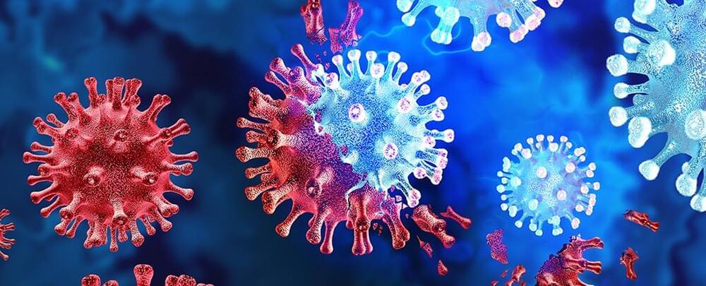
The Covid-19 pandemic continues to be a terrible plight on the world. But if there’s any silver lining in what has happened, the deadly worldwide plague has brought about advancements in medicine created to fight it that may have transformational impacts well past the pandemic.
In a conversation during CES 2023, one of the world’s eminent tech gatherings, Moderna CEO Stéphane Bancel discussed the potential to utilize mRNA technology at the basis of groundbreaking Covid vaccines, developed with unprecedented speed, to create personalized cancer treatments.
Yuuji/iStock.
In a conversation during CES 2023, one of the world’s eminent tech gatherings, Moderna CEO Stéphane Bancel discussed the potential to utilize mRNA technology at the basis of groundbreaking Covid vaccines, developed with unprecedented speed, to create personalized cancer treatments. The conversation “Personalized Medicine: The Future of Cancer Treatment is Not One-Size Fits All” was led by Stephen Klasko, executive in residence at General Catalyst and advisor at Stel Life.
Researchers at Princeton’s Department of Chemistry discovered the first known de novo protein that catalyzes, or drives, the synthesis of quantum dots.
Nature uses 20 canonical amino acids.
<div class=””> <div class=””><br />Amino acids are a set of organic compounds used to build proteins. There are about 500 naturally occurring known amino acids, though only 20 appear in the genetic code. Proteins consist of one or more chains of amino acids called polypeptides. The sequence of the amino acid chain causes the polypeptide to fold into a shape that is biologically active. The amino acid sequences of proteins are encoded in the genes. Nine proteinogenic amino acids are called “essential” for humans because they cannot be produced from other compounds by the human body and so must be taken in as food.<br /></div> </div>
Newspaper headlines from the U.S. to the U.K. and most places in between highlight the surge in sick patients suffering from respiratory viruses. The so-called “tripledemic” of lung infections including respiratory synclinal virus (RSV), influenza (flu) and COVID-19 (coronavirus) is likely to last throughout the winter season. This explosion of infections requires more treatment options to support overloaded hospitals and overworked medics as they restore people’s health.
It has been known for a long time that intubation of an infant with any lung condition, or even an adult with severe COVID-19 using either ventilation or extra-corporeal membrane oxygenation (ECMO), comes with risks and side effects that could cause permanent damage not limited to the lungs. However, hypoxia, which means oxygen deficiency, is a medical emergency that is a common complication of severe lung infections. If not treated, it can lead to severe disability and even death.
A type of freshwater plankton has become the first organism seen thriving on a diet of viruses, according to a new study by researchers from the University of Nebraska-Lincoln in the US.
Viruses are often consumed incidentally by a range wide of organisms, and may even season the diets of certain marine protists. But to qualify as a true step in the food chain – described as virovory – viruses ought to contribute a significant amount of energy or nutrients to their consumer.
The microbe Halteria is a common genus of protist known to flit about as its hair-like cilia propel it through the water. Not only did laboratory samples of the ciliate consume chloroviruses added to its environment, the giant virus fueled Halteria’s growth and increased its population size.
Point-of-care diagnoses occur when a doctor can quickly diagnose a patient during an examination without sending biological samples to a laboratory or consulting with other specialists. The stress and anxiety that can come with waiting for the results of medical testing, and the cost associated with in-depth laboratory testing, make point-of-care testing a gold standard. However, point-of-care diagnostics remain rare in oncology.
Research shows that women suspected of endometrial cancer experience stress and anxiety while waiting for a confirmed diagnosis. While the procedures and waiting time associated with endometrial cancer diagnoses vary, endometrial biopsies can take weeks to return results. Of added concern, since most endometrial cancers require surgical intervention, usually a hysterectomy, delays in diagnosis lead to delays in treatment. Indeed, these surgical delays can negatively impact survival. Thus, rapid endometrial cancer diagnostic strategies would significantly improve patient care.
To address the need for new, more efficient endometrial cancer diagnostics, a group of researchers initiated a study to assess the ability of an intelligent knife (iKnife) to identify malignancy in endometrial tissue biopsies. The researchers published the results of the study in the journal Cancers.