Toggle light / dark theme

Heterogeneity in immune cell abnormalities in patients with systemic sclerosis

“We know that immune dysregulation causes vascular damage and tissue fibrosis in systemic sclerosis,” says the lead author. “However, it remains unclear why skin symptoms and the level of organ involvement differ from patient to patient.”

To explore this, the researchers took blood and tissue samples from patients with systemic sclerosis and analyzed them cell by cell, looking for differences in gene expression. Additionally, proteins on the cell surface were examined to identify biomarkers of disease, which are useful for identifying and treating diseases in earlier stages.

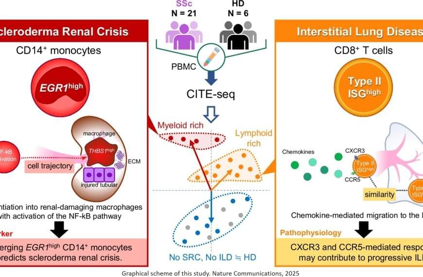
“The results were intriguing,” explains the senior author. “We identified a specific subset of immune cells, the EGR1-expressing subpopulation of CD14+ monocytes, that were clearly associated with scleroderma renal crisis, a serious kidney complication in patients with systemic sclerosis.”

While immune cells usually help the body fight infection and disease, in certain cases they can be inappropriately activated through gene expression. In this instance, CD14+ monocytes differentiated – or transformed – into destructive macrophages, which can further promote inflammation near the kidneys and contribute to the thickening and scarring of internal organs.

In addition, the researchers found that CD8+ T cells with a type II interferon signature, which makes the immune cells particularly aggressive and inflammatory, were linked to progressive interstitial lung disease. EGR1-expressing CD14+ monocytes and these peculiar CD8+ T cells are likely to accumulate in the kidney or the lung, respectively, and produce or recruit other factors that contribute to disease progression.


Treating rare diseases can be complicated at the best of times, and it gets even more complicated when different patients with the same disease exhibit different symptoms. Now, researchers have reported a cellular signature that might explain why some patients with autoimmune disease are stable while others face life-threatening complications.

Team discovers how tiny parts of cells stay organized, adding new insights for blocking cancer growth

A team of international researchers led by scientists at City of Hope provides the most thorough account yet of an elusive target for cancer treatment. Published in Science Advances, the study suggests a complex signaling process involving paxillin, a focal adhesion protein that acts as a hub to connect with other proteins, may be vulnerable to therapy despite its fluid state.

“Disrupting the interaction of paxillin with bears direct relevance in ,” said Ravi Salgia, M.D., Ph.D., the Arthur & Rosalie Kaplan Chair in Medical Oncology at City of Hope’s comprehensive cancer center. “This can lead to precision therapeutics targeting a specific paxillin function that is dominant in cancer cells, but less prevalent in healthy cells.”

The research adds important new details on a hard-to-characterize network of cellular proteins. Dr. Salgia and his team looked closely at paxillin, which prompts cells to change in response to the environment. This helps to evolve and evade detection, while also causing resistance to treatment. Dr. Salgia and his team have been working on elucidating the function of paxillin for over three decades. He and his colleagues were the first to clone the full-length human gene in 1995 at Harvard.

Scientists uncover kidney-to-brain route for Parkinson’s-related protein spread

A groundbreaking study suggests that Parkinson’s disease may begin in the kidneys, where a toxic protein builds up and travels to the brain. This discovery could reshape our understanding of the disease’s origins and risk factors.

Atherosclerotic blood vessel cells grow similar to tumors, study reveals

Researchers from the University of Southern Denmark and Odense University Hospital have studied tissue from patients with atherosclerosis. They found that many of the cells in the diseased tissue carried the same genetic alteration and appeared to originate from a single ancestral cell that had divided repeatedly—a pattern otherwise associated with tumor biology.

In several patients, a large proportion of the cells were derived from one single mutated cell that had undergone many rounds of cell division.

“It’s striking how many cells in the tissue share the exact same . In several samples, more than 10% of the cells—hundreds of thousands cells—carried the same alteration. It’s difficult to interpret this as anything other than all these cells originating from a shared ancestral cell that, at some point during disease development, acquired the mutation,” says Lasse Bach Steffensen, Associate Professor at the Department of Molecular Medicine at the University of Southern Denmark.

Scientists Crack the 500-Million-Year-Old Code That Controls Your Immune System

A collaborative team from Penn Medicine and Penn Engineering has uncovered the mathematical principles behind a 500-million-year-old protein network that determines whether foreign materials are recognized as friend or foe. How does your body tell the difference between friendly visitors, like me

Alzheimer’s: Bacteria that cause stomach ulcers may also protect brain

Every three seconds, someone in the world develops dementia. Alzheimer’s disease is the most common form of dementia, accounting for between 60% and 70% of all cases.

Although scientists have made significant progress in understanding the disease, there’s still no cure. That’s partly because Alzheimer’s disease has multiple causes—many of which are still not fully understood.

Two proteins which are widely believed to play central roles in Alzheimer’s disease are amyloid-beta and tau. Amyloid-beta forms sticky plaques on the outside of brain cells. This disrupts communication between neurons. Tau accumulates inside brain cells, where it twists into tangles. This ultimately leads to cell death. These plaques and tangles are the hallmark features of Alzheimer’s disease.

Scientists find cellular brain changes tied to PTSD

The human brain is made up of billions of interconnected cells that are constantly talking to each other. A new study published in Nature zooms in to the single-cell level to see how this cellular communication may be going wrong in brains affected by post-traumatic stress disorder (PTSD).

Until recently, researchers did not have the technology to study within individual cells. But now that it’s available, a team led by Matthew Girgenti, Ph.D., assistant professor of psychiatry at Yale School of Medicine, has been analyzing to uncover genetic variants that might be associated with psychiatric diseases such as (MDD) and PTSD.

Their latest study is one of the first to examine a major psychiatric disorder, PTSD, at the single-cell level. For years, doctors have been prescribing antidepressants to treat the condition because there are currently no drugs specifically designed for PTSD. Girgenti hopes that identifying novel molecular signatures associated with the psychiatric disease can help researchers learn how to develop new drugs or repurpose existing ones to treat it more effectively.

Scientists demonstrate unconditional exponential quantum scaling advantage using two 127-qubit computers

Quantum computers have the potential to speed up computation, help design new medicines, break codes, and discover exotic new materials—but that’s only when they are truly functional.

One key thing that gets in the way: noise or the errors that are produced during computations on a quantum machine—which in fact makes them less powerful than —until recently.

Daniel Lidar, holder of the Viterbi Professorship in Engineering and Professor of Electrical & Computer Engineering at the USC Viterbi School of Engineering, has been iterating on , and in a new study along with collaborators at USC and Johns Hopkins, has been able to demonstrate a quantum exponential scaling advantage, using two 127-qubit IBM Quantum Eagle processor-powered quantum computers, over the cloud.

/* */