Toggle light / dark theme

Full coverage of the coronavirus outbreak

Germany started cautiously easing restrictions earlier this month. But the spread of more infectious variants of the virus has pushed up cases, prompting concerns that hospitals could soon be overstretched without further curbs.

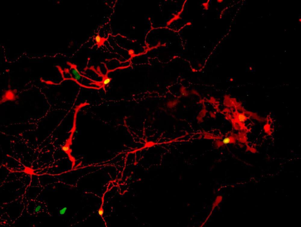
The research team, led by Todd Lencz, PhD, with Itsik Pe’er, PhD, Tom Maniatis, PhD, and Erin Flaherty, PhD, of Columbia University, carried out a genetic study identifying a single letter change in the DNA code in the PCDHA3 gene that is associated with schizophrenia. The affected gene makes a type of protein called a protocadherin, which generates a cell surface “barcode” required for neurons to recognize, and communicate with, other neurons. They found that the PCDHA3 variant blocks this normal protocadherin function.

The discovery was made possible by the special genetic characteristics of the samples studied by Lencz’s team—patients with schizophrenia and healthy volunteers drawn from the Ashkenazi Jewish population. The Ashkenazi Jewish population represents an important population for study based on its unique history. Just a few hundred individuals who migrated to Eastern Europe less than 1000 years ago are the ancestors of nearly 10 million Ashkenazi Jews today. This lineage, combined with a tradition of marriage within the community, has resulted in a more uniform genetic background in which to identify disease-related variants.

“In addition to our primary findings regarding PCDHA3 and related genes, we were able— due to the unique characteristics of the Ashkenazi population—to replicate several prior findings in schizophrenia despite relatively small sample sizes,” said Lencz, professor in the Institute of Behavioral Science at the Feinstein Institutes. “In our study, we demonstrated this population represents a smart, cost-effective strategy for identifying disease-related genes. Our findings allow us to zero in on a novel aspect of brain development and function in our quest to develop new treatments for schizophrenia.”

It is well established that rare, damaging genetic variants with strong effects contribute to autism. Although individually rare, these variants are collectively common: Clinical genetic testing identifies them in at least 25 percent of autistic people. Studies of these variants have implicated more than 100 genes — and counting — in autism.

Identifying these genes is important — not only for clinical care, but also for advancing our understanding of the neural circuits and processes involved in autism or in its core traits. It creates the opportunity to develop therapies targeted to specific molecular diagnoses. And as we learn more about these genes and the consequences of variants that disrupt their function, we have the potential to better understand the mechanisms underlying cases of autism in which a definitive genetic diagnosis cannot yet be made.

But the genetic findings in people with autism are not unique; deleterious variants in the same genes are also implicated in other neurodevelopmental conditions, such as intellectual disability, epilepsy, attention deficit hyperactivity disorder and schizophrenia. Specific genes and variants do not map neatly onto categorical clinical diagnoses or the core cognitive and behavioral traits that define them. In fact, there is not yet a single example of a gene that, when mutated, increases the likelihood of autism but not of other neurodevelopmental conditions.

Researchers at Chalmers University of Technology, Gothenburg, Sweden, have developed a novel type of thermometer that can simply and quickly measure temperatures during quantum calculations with extremely high accuracy. The breakthrough provides a benchmarking tool for quantum computing of great value—and opens up for experiments in the exciting field of quantum thermodynamics.

Key components in quantum computers are coaxial cables and waveguides—structures that guide waveforms and act as the vital connection between the and the classical electronics that control it. Microwave pulses travel along the waveguides to the quantum processor, and are cooled down to extremely along the way. The also attenuates and filters the pulses, enabling the extremely sensitive quantum computer to work with stable quantum states.

In order to maximize control over this mechanism, the researchers need to be sure that these waveguides are not carrying noise due to thermal motion of electrons on top of the pulses that they send. In other words, they have to measure the temperature of the electromagnetic fields at the cold end of the microwave waveguides, the point where the controlling pulses are delivered to the computer’s qubits. Working at the lowest possible temperature minimizes the risk of introducing errors in the qubits.

Humans have the innate ability to store important information in their mind for short periods of time, a capability known as short-term memory. Over the past few decades, numerous neuroscientists have tried to understand how neural circuits store short-term memories, as this could lead to approaches to assist individuals whose memory is failing and help to devise memory enhancing interventions.

Researchers at Stanford and the Janelia Research Campus, Howard Hughes Medical Institute have recently identified neural circuit motifs involved in how humans store short-term memories. Their findings, published in Nature Neuroscience, suggest that -related contain recurrently connected modules that independently maintain selective and continuous activity.

“Short-term memories are of approximately 10 seconds or so, for example, if you needed to remember a while you looked for a pen to write the number,” Kayvon Daie, one of the researchers who carried out the study, told Medical Xpress. “Individual , however, are very forgetful, as they can only remember their inputs for about 10 milliseconds. It has been hypothesized that if two forgetful neurons were connected to each other, they could continuously remind each other of what they were supposed to remember so that the circuit can now hold information for many seconds.”

For decades, people undergoing radiotherapy, which is used to treat cancer, have reported a bizarre phenomenon: Seeing flashes of light in their eyes, even when their eyes are closed.

Patients documented in the medical literature have described a ‘‘ray of blue light” and ‘‘seeing a blue neon light”, sometimes accompanied by a “white smell” during the delivery of radiation, lasting for a fraction of a second. There have been several theories for why this could be happening, including retinal pigments inside patients’ eyes being stimulated during the therapy, or that Cherenkov light or Cherenkov radiation – the same effect that makes nuclear reactors glow blue when they’re underwater – is produced inside the eyeball itself.

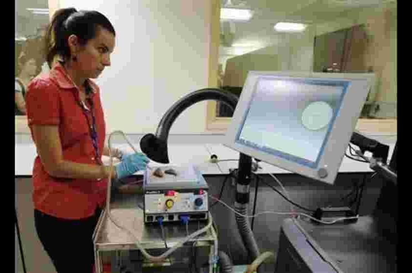
Now scientists have captured this strange light for the first time, producing the first photographic evidence that the phenomenon is in fact Cherenkov light.

“Previously reported detection of plant biomagnetism, which established the existence of measurable magnetic activity in the plant kingdom, was carried out using superconducting-quantum-interference-device (SQUID) magnetometers1, 5, 16. Atomic magnetometers are arguably more attractive for biological applications, since, unlike SQUIDs34, 35, they are non-cryogenic and can be miniaturized to optimize spatial resolution of measured biological features14, 15, 36. In the future, the SNR of magnetic measurements in plants will benefit from optimizing the low-frequency stability and sensitivity of atomic magnetometers. Just as noninvasive magnetic techniques have become essential tools for medical diagnostics of the human brain and body, this noninvasive technique could also be useful in the future for crop-plant diagnostics—by measuring the electromagnetic response of plants facing such challenges as sudden temperature change, herbivore attack, and chemical exposure.”


Upon stimulation, plants elicit electrical signals that can travel within a cellular network analogous to the animal nervous system. It is well-known that in the human brain, voltage changes in certain regions result from concerted electrical activity which, in the form of action potentials (APs), travels within nerve-cell arrays. Electro-and magnetophysiological techniques like electroencephalography, magnetoencephalography, and magnetic resonance imaging are used to record this activity and to diagnose disorders. Here we demonstrate that APs in a multicellular plant system produce measurable magnetic fields. Using atomic optically pumped magnetometers, biomagnetism associated with electrical activity in the carnivorous Venus flytrap, Dionaea muscipula, was recorded. Action potentials were induced by heat stimulation and detected both electrically and magnetically.

Imagine not a white, but a green Arctic, with woody shrubs as far north as the Canadian coast of the Arctic Ocean. This is what the northernmost region of North America looked like about 125000 years ago, during the last interglacial period, finds new research from CU Boulder.

Researchers analyzed plant DNA more than 100000 years old retrieved from lake sediment in the Arctic (the oldest DNA in lake sediment analyzed in a publication to date) and found evidence of a shrub native to northern Canadian ecosystems 250 miles (400 km) farther north than its current range.

As the Arctic warms much faster than everywhere else on the planet in response to climate change, the findings, published this week in the Proceedings of the National Academy of Sciences, may not only be a glimpse of the past but a snapshot of our potential future.