Toggle light / dark theme

New insight into the generation of new neurons in the adult brain

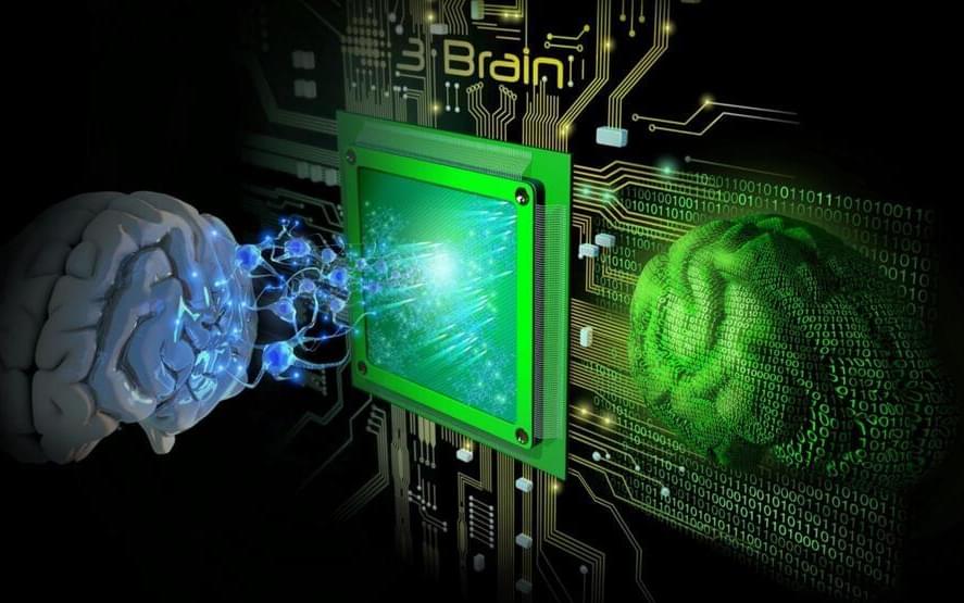
Researchers at Sahlgrenska Academy at the University of Gothenburg, Sweden, in collaboration with research groups in Finland, Canada and Slovenia, have discovered a novel and unexpected function of nestin, the best-known marker of neural stem cells.
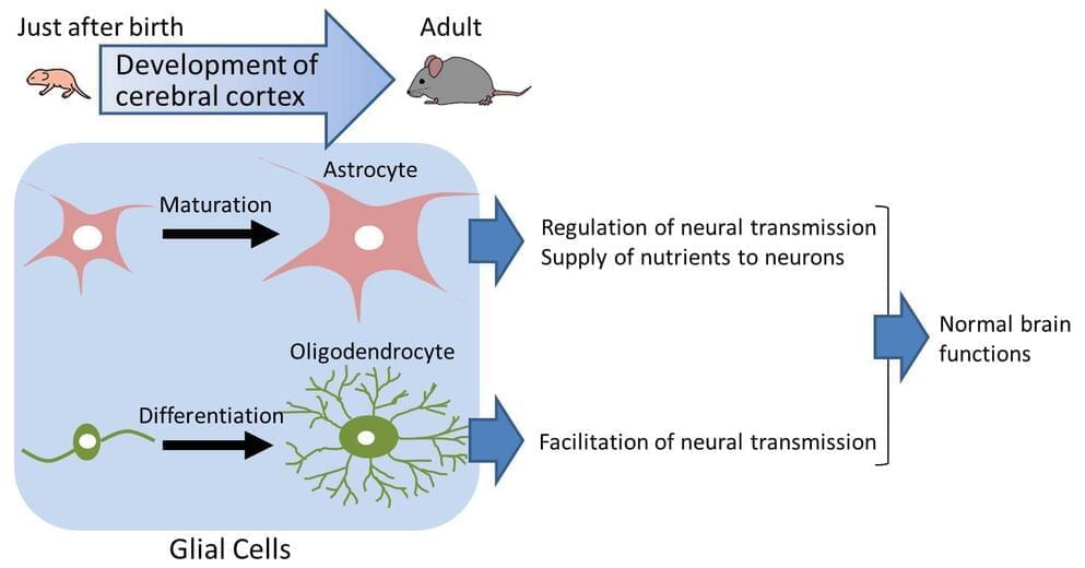
In the , the three main cell types, neurons, astrocytes and oligodendrocytes, are generated from neural stem . In some such as the hippocampus, the region involved in learning and memory, new neurons are being added to the existing neuronal circuitry even in adulthood, when severe restriction of neuronal differentiation occurs.

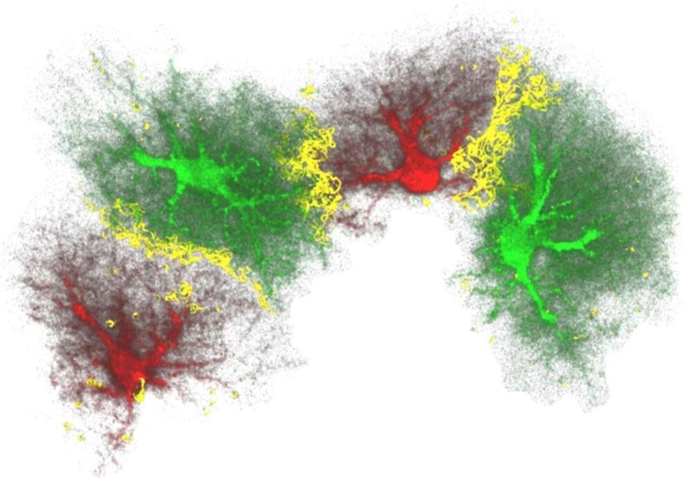
Using mice deficient in nestin, a protein that is a component of the part of the cytoskeleton known as or nanofilaments, the research team led by Prof. Milos Pekny showed that nestin produced in astrocytes has an important role in inhibiting neuronal differentiation. They linked this regulatory function of nestin to the Notch signaling from astrocytes to neighboring neural stem cells. Thus, surprisingly, nestin does not control the generation of neurons by acting within neural stem cells, but indirectly by regulating the neurogenesis-inhibitory Notch signals that receive from astrocytes, important constituents of the neurogenic niche.

CD38 gene is identified to be important in postnatal development of the cerebral cortex

The brain consists of neurons and glial cells. The developmental abnormality of glial cells causes various diseases and aberrant cerebral cortex development. CD38 gene knockout is shown to cause aberrant development of glial cells, especially astrocytes and oligodendrocytes. The CD38 gene is known to be involved in cerebral cortex development. The present study suggests the importance of glial cells for cerebral cortex development.

It is essential for that both neurons and develop in a normal manner not only during fetal but also postnatal stages. In the postnatal brain, neurons extend long protrusions (axons and dendrites) to form complex networks for information exchange. On the other hand, glial are thought to support network formation of neurons, to regulate transmission of information, and to help survival of neurons. It is known that more than 50 percent of total cells in the brain are glial cells, three times more than neurons in number. It is also known that in the human brain has far more glial cells than the brains of rodents or primates. This indicates that for the higher functions of the brain, glial cells are of particular importance.

In the past, research on developmental disorders of the brain focused on neurons. Recently, however, research has focused on the abnormality of glial cells. There remain a number of unsolved problems concerning the mechanism of glial cell development in the postnatal brain and the relationship of glial cell abnormalities and developmental disorders of the brain.

Adding human glial cells to mice brains found to improve memory and cognition

(Medical Xpress)—A team of researchers working at the University of Rochester in New York, has found that injecting glial cells into a mouse brain caused an improvement in both memory and cognition in the mouse. In their paper published in The Journal of Neuroscience, the team explains how they injected the test mice and then tested them afterwards to see what impact it had on their abilities.

Injecting human into mice brains appears to be the stuff of horror films, but in this case, it wasn’t really what it might have seemed. Glial cells are precursors to other cells—in this case, they develop into astrocytes, which are technically, brain cells. But, the important distinction here is that they are not neurons, which means they are not involved in thinking—instead they are involved in memory retention and help with housekeeping tasks.

Last year, the team injected mature into mice brains and reported improvements in ability by the mice—this time they went further, injecting progenitor precursor glial cells, which allows for development of more astrocytes. The team injected just 300,000 of the cells (from donated human embryos) and found just 12 months later that they had multiplied to grow to 12 million, completely displacing the original mouse astrocytes. It appeared, the team reported, that the cell growth only stopped when it reached the physical confines of the skull. They also note that it was interesting that the glial could thrive in such an environment considering that astrocytess in people are 10 to 20 times as big as those in mice and they carry 100 times as many tendrils. Testing the mice showed that their memory was far superior to normal mice and they had improved cognition as well.

‘Synthetic’ mouse embryo with brain and beating heart grown from stem cells

Researchers from the University of Cambridge and Caltech have created model mouse embryos from stem cells—the body’s master cells, which can develop into almost any cell type in the body—that have beating hearts, as well as the foundations for a brain and all of the other organs in the mouse body.

The results are the culmination of more than a decade of research, and they could help researchers understand why some embryos fail while others go on to develop into a fetus as part of a healthy pregnancy. Additionally, the results could be used to guide repair and development of synthetic human organs for transplantation.

The research was conducted in the laboratory of Magdalena Zernicka-Goetz, Bren Professor of Biology and Biological Engineering at Caltech. Zernicka-Goetz is also a professor of mammalian development and in Cambridge’s Department of Physiology, Development and Neuroscience. A paper describing the breakthrough appears in the journal Nature on August 25.

Researchers build embryo-like structures from human stem cells

Research on human embryos is vital to understanding the earliest stages of human development. Currently, this research is conducted on surplus embryos willingly donated by individuals who have undergone in vitro fertilization. Nevertheless, this research is limited by the availability of embryos and strict international ethical time limits on how long an embryo is allowed to develop in the laboratory (14 days maximum.)

Now, Caltech researchers have created embryo-like structures out of human stem cells. In contrast to natural embryos that are formed by a combination of sperm and egg, these structures are formed by combining so-called , which have the ability to develop into specialized types of cells. Though these embryo-like structures have some key differences from real embryos, the technology to create them will be critical in answering open questions about without the need for donated embryos.

The research was conducted in the laboratory of Magdalena Zernicka-Goetz, Bren Professor of Biology and Biological Engineering at Caltech, and is described in a paper appearing in the journal Nature Communications on September 21.

Scientists use stem cells to create synthetic mouse embryos

Scientists have created “synthetic” mouse embryos from stem cells without a dad’s sperm or a mom’s egg or womb.

The lab-created embryos mirror a natural mouse embryo up to 8 ½ days after fertilization, containing the same structures, including one like a beating heart.

In the near term, researchers hope to use these so-called embryoids to better understand early stages of development and study mechanisms behind disease without the need for as many lab animals. The feat could also lay the foundation for creating synthetic human embryos for research in the future.

Efficient AI technology for MRI data analysis

An algorithm developed by researchers from Helmholtz Munich, the Technical University of Munich (TUM) and its University Hospital rechts der Isar, the University Hospital Bonn (UKB) and the University of Bonn is able to learn independently across different medical institutions. The key feature is that it is self-learning, meaning it does not require extensive, time-consuming findings or markings by radiologists in the MRI images.

This federated was trained on more than 1,500 MRI scans of healthy study participants from four institutions while maintaining data privacy. The algorithm then was used to analyze more than 500 patient MRI scans to detect diseases such as multiple sclerosis, vascular disease, and various forms of brain tumors that the algorithm had never seen before. This opens up new possibilities for developing efficient AI-based federated algorithms that learn autonomously while protecting privacy. The study has now been published in the journal Nature Machine Intelligence.

Health care is currently being revolutionized by artificial intelligence. With precise AI solutions, doctors can be supported in diagnosis. However, such algorithms require a considerable amount of data and the associated radiological specialist findings for training. The creation of such a large, central database, however, places special demands on . Additionally, the creation of the findings and annotations, for example the marking of tumors in an MRI image, is very time-consuming.

Nontoxic material found to be ultra-strong solar energy harvester

Solar cells are vital for the green energy transition. They can be used not only on rooftops and solar farms but also for powering autonomous vehicles, such as planes and satellites. However, photovoltaic solar cells are currently heavy and bulky, making them difficult to transport to remote locations off-grid, where they are much needed.

In a collaboration led by Imperial College London, alongside researchers from Cambridge, UCL, Oxford, Helmholtz-Zentrum Berlin in Germany, and others, researchers have produced that can absorb comparable levels of sunlight as conventional silicon , but with 10,000 times lower thickness.

The material is sodium bismuth sulfide (NaBiS2), which is grown as nanocrystals and deposited from solution to make films 30 nanometers in thickness. NaBiS2 is comprised of nontoxic elements that are sufficiently abundant in the earth’s crust for use commercially. For example, bismuth-based compounds are used as a nontoxic lead replacement in solder, or in over-the-counter stomach medicine.