Toggle light / dark theme

You might not recognize the term NSAID, which stands for non-steroidal anti-inflammatory drugs, but you are undoubtedly familiar with this class of medications which are commonly used for headaches, pain relief, and to reduce fever. You’ll also recognize the common types of NSAIDs including aspirin, ibuprofen, and naproxen, as well as the brand names such as Bayer, St. Joseph, Advil, Motrin, and Aleve.

Even though they are often used to treat pain and inflammation, different NSAIDs can have surprising and inexplicable effects on many diseases. New research reveals a previously unknown process by which some NSAIDs affect the body. The findings may explain the varying effects of different NSAIDs and also point to entirely new applications for these drugs.

Since these medications are so widely used, it is really important to fully understand how they affect the body.

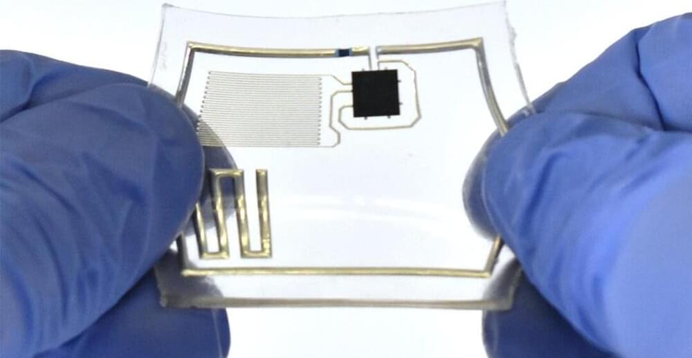
An Apple Watch owner has created a complication and watchOS app that works with a glucose monitor, so they can keep track of their blood glucose level from their wrist.

Numerous rumors have claimed Apple is actively working on some form of glucose monitoring sensor for the Apple Watch, but has so far yet to add it to the wearable device. In the case of one Apple Watch owner, they managed to hack together their own solution.

The project, outlined by Harley Turan, effectively takes the data from a continuous glucose monitoring system and imports and interprets it in a way that it can be viewed on an Apple Watch. In doing so, the project creates a reasonably low-cost solution for the problem.

Aqueous droplet formation by liquid-liquid phase separation (or coacervation) in macromolecules is a hot topic in life sciences research. Of these various macromolecules that form droplets, DNA is quite interesting because it is predictable and programmable, which are qualities useful in nanotechnology. Recently, the programmability of DNA was used to construct and regulate DNA droplets formed by coacervation of sequence designed DNAs.

A group of scientists at Tokyo University of Technology (Tokyo Tech) led by Prof. Masahiro Takinoue has developed a computational DNA droplet with the ability to recognize specific combinations of chemically synthesized microRNAs (miRNAs) that act as biomarkers of tumors. Using these miRNAs as molecular input, the can give a DNA logic computing output through physical DNA droplet phase separation. Prof. Takinoue explains the need for such studies, “The applications of DNA droplets have been reported in cell-inspired microcompartments. Even though regulate their functions by combining biosensing with molecular logical computation, no literature is available on integration of DNA droplet with molecular computing.” Their findings were published in Advanced Functional Materials.

Developing this DNA droplet required a series of experiments. First, they designed three types of Y-shaped DNA nanostructures called Y-motifs A, B, and C with 3 sticky ends to make A, B, and C DNA droplets. Typically, similar droplets band together automatically while to join dissimilar droplets a special “linker” molecule is required. So, they used linker molecules to join the A droplet with the B and C droplets; these linker molecules were called AB and AC linkers, respectively.

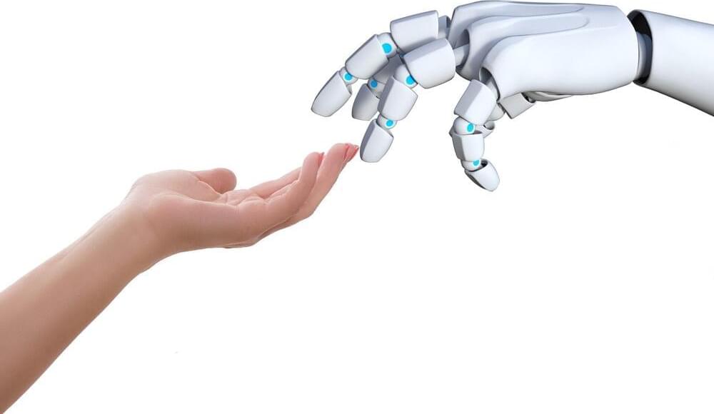
Transhumanism is a movement that aims to address – or end – what Bohan calls the “tragedies of reality”: ageing, sickness and involuntary death. It is, she writes, “a philosophy and a project that aims to make us more than human”.

Whether we recognise or understand it, that project has already begun, she says, and it will transform our world – and minds and bodies – within our lifetimes. Not only is it happening, she says, but this transition is necessary if humanity is to survive in perpetuity.

For Bohan, it is no great to leap to imagine that a baby born in 2030 may have its entire genome mapped at birth, that data uploaded to a central health record and cross-referenced at any medical appointment throughout its life. It is no great stretch to think that AI will become the most powerful intellectual force of the century. That human consciousness might be transferred from our “meat sacks” (bodies) into a technological sphere. That the rise of AI and automation might render great swathes of human labour redundant, and that maybe – if we get it right – that could leave more time for leisure, big thinking, meditation, connection.

Circa 2019 immortality in the human brain 🧠


Brain injuries causing chronic sensory or motor deficit, such as stroke, are among the leading causes of disability worldwide, according to the World Health Organization; furthermore, they carry heavy social and economic burdens due to decreased quality of life and need of assistance. Given the limited effectiveness of rehabilitation, novel therapeutic strategies are required to enhance functional recovery. Since cell-based approaches have emerged as an intriguing and promising strategy to promote brain repair, many efforts have been made to study the functional integration of neurons derived from pluripotent stem cells (PSCs), or fetal neurons, after grafting into the damaged host tissue. PSCs hold great promises for their clinical applications, such as cellular replacement of damaged neural tissues with autologous neurons. They also offer the possibility to create in vitro models to assess the efficacy of drugs and therapies. Notwithstanding these potential applications, PSC-derived transplanted neurons have to match the precise sub-type, positional and functional identity of the lesioned neural tissue. Thus, the requirement of highly specific and efficient differentiation protocols of PSCs in neurons with appropriate neural identity constitutes the main challenge limiting the clinical use of stem cells in the near future. In this Review, we discuss the recent advances in the derivation of telencephalic (cortical and hippocampal) neurons from PSCs, assessing specificity and efficiency of the differentiation protocols, with particular emphasis on the genetic and molecular characterization of PSC-derived neurons. Second, we address the remaining challenges for cellular replacement therapies in cortical brain injuries, focusing on electrophysiological properties, functional integration and therapeutic effects of the transplanted neurons.

Brain injuries represent a large variety of disabling pathologies. They may originate from different causes and affect distinct brain locations, leading to an enormous multiplicity of various symptoms ranging from cognitive deficits to sensorimotor disabilities. They can also result in secondary disturbances, such as epileptic foci, which occur within the lesioned and perilesional tissues (Herman, 2002). Indeed, frequently a secondary functional damage can take place in a region distant from the first insult (e.g., the hippocampus after traumatic brain injury), providing an explanation for cognitive and memory deficits arising after a brain lesion (Girgis et al., 2016). Brain injuries can have traumatic or non-traumatic etiologies, including focal brain lesions, anoxia, tumors, aneurysms, vascular malformations, encephalitis, meningitis and stroke (Teasell et al., 2007). In particular, stroke covers a vast majority of acquired brain lesions.