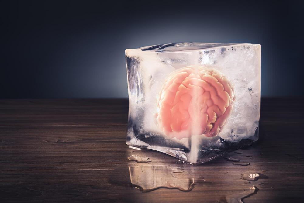
Toggle light / dark theme

The Brain Chemical Involved in Consciousness

So how do we help these people? The brain is more than just a congregation of different areas. Brain cells also rely on a number of chemicals to communicate with other cells, enabling a number of brain functions. Before our study, there was already some evidence that dopamine, well known for its role in reward, also plays a role in disorders of consciousness.

For example, one study showed that dopamine release in the brain is impaired in minimally conscious patients. Moreover, a number of small-scale studies have shown that patients’ consciousness can improve by giving them drugs that act through dopamine.

Flexible electronics have enabled the design of sensors, actuators, microfluidics and electronics on flexible, conformal and/or stretchable sublayers for wearable, implantable or ingestible applications. However, these devices have very different mechanical and biological properties when compared to human tissue and thus cannot be integrated with the human body.

A team of researchers at Texas A&M University has developed a new class of biomaterial inks that mimic native characteristics of highly conductive , much like skin, which are essential for the ink to be used in 3D printing.

This biomaterial ink leverages a new class of 2D nanomaterials known as molybdenum disulfide (MoS2). The thin-layered structure of MoS2 contains defect centers to make it chemically active and, combined with modified gelatin to obtain a flexible hydrogel, comparable to the structure of Jell-O.

And these miniaturized brains could save regular-sized brains.

Electroencephalography (EEG) caps are medical devices doctors use to diagnose brain disorders like epilepsy and seizures in patients. In the past decade, scientists have created 3D mini-brains called brain organoids from human-derived stem cells that mimic some aspects of brain development. A team of researchers at John Hopkins University has recently developed the world’s smallest EEG caps to study these more efficiently. The micro EEG caps can be used on a brain organoid the size of a pen dot.

Brain organoids can mimic some key features of the human brain. Scientists create them to understand the human brain’s development process and the factors leading to various neural disorders. Moreover, such mini-brains can also be used to perform experiments that researchers would have to otherwise perform on a real brain. Thus, eliminating the need to conduct tests on live human and animal subjects. has long been the stuff of science fiction but now mind-reading machines may actually be here and they may not be invasive. Researchers from the Russian corporation Neurobotics and the Moscow Institute of Physics and Technology have found a way to visualize a person’s brain activity as actual images without the use of invasive brain implants.

For many processes important for life such as cell division, cell migration, and the development of organs, the spatially and temporally correct formation of biological patterns is essential. To understand these processes, the principal task consists not in explaining how patterns form out of a homogeneous initial condition, but in explaining how simple patterns change into increasingly complex ones. Illuminating the mechanisms of this complex self-organization on various spatial and temporal scales is a key challenge for science.

So-called “coarse-graining” techniques allow such multiscale systems to be simplified, such that they can be described with a reduced model at large length and time scales. “The price you pay for coarse-graining, however, is that important information about the patterns on small scales—like the pattern type—is lost. But the thing is that these patterns play a decisive role in . To give one example, they control important cellular processes,” explains Laeschkir Würthner, member of the team led by LMU physicist Prof. Erwin Frey and lead author of a new study published in the Proceedings of the National Academy of Sciences that overcomes this issue.

In collaboration with the research group of Prof. Cees Dekker (TU Delft), Frey’s team has developed a new coarse-graining approach for so-called mass-conserving reaction-diffusion systems, in which the large-scale analysis of the total densities of the particles involved enables the prediction of patterns on small scales.

Cells use selective autophagy or self-degradation of undesired proteins to maintain cellular homeostasis (i.e., a state of balance). This process is controlled by autophagy receptors, which mediate the selection of a target protein that is subsequently “cleared.”

Tau proteins, which play a crucial role in the internal architecture of neurons in the brain, abnormally accumulate within neurons in disorders such as dementia and Alzheimer’s.

Alzheimer’s disease is a disease that attacks the brain, causing a decline in mental ability that worsens over time. It is the most common form of dementia and accounts for 60 to 80 percent of dementia cases. There is no current cure for Alzheimer’s disease, but there are medications that can help ease the symptoms.

In the 1980s, biologist Dr Michael Rose started to selectively breed Drosophila fruit flies for increased longevity. Today, the descendants of the original Methuselah flies are held by biotech firm Genescient Corporation and live 4.5 times longer than normal fruit flies.

The flies’ increased lifespan is explained by a significant number of systemic genetic changes — but how many of these variations represent lessons that can be used to design longevity therapies for humans? Dr. Ben Goertzel and his bio-AI colleagues at SingularityNET and Rejuve. AI are betting the answer is quite a few.

SingularityNET and Rejuve. AI have launched a partnership with Genescient to apply advanced machine learning and machine reasoning methods to transfer insights gained from the Methuselah fly genome to the human genome. The goal is to acquire new information regarding gene therapies, drugs or nutraceutical regimens for prolonging healthy human life.

Damage to the ends of your chromosomes can create “zombie cells” that are still alive but can’t function, according to our recently published study in Nature Structural and Molecular Biology.

When cells prepare to divide, their DNA is tightly wound around proteins to form chromosomes that provide structure and support for genetic material. At the ends of these chromosomes are repetitive stretches of DNA called telomeres that form a protective cap to prevent damage to the genetic material.

However, telomeres shorten each time a cell divides. This means that as cells divide more and more as you age, your telomeres become increasingly shorter and more likely to lose their ability to protect your DNA.

Scientists experimenting with next-generation plastics at Finland’s University of Turku have developed a form of the material with some impressive capabilities, most notably an ability to quickly break down after use. The eco-friendly “supramolecular” plastic is therefore highly recyclable and, with careful tuning of its water content, can be turned into an adhesive or even instantly self-heal when damaged.

The reason conventional plastics persist in the environment for so long is the incredibly strong chemical connections between the monomers within them. These particles link up to form polymers through what are known as covalent bonds, but scientists hope to fashion more environmentally forms of the material based on non-covalent bonds instead.

These weaker connections are better suited to degradation and recycling of the material, but do come at a cost in terms of mechanical performance. We have looked at some interesting examples of these “supramolecular” materials in the form of hybrid polymers for drug delivery, self-assembling plastics and adhesives that work at extreme temperatures.