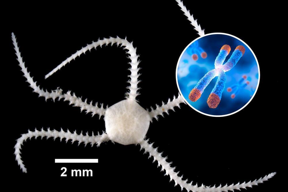
Chromosomes are threadlike structures composed entirely of DNA that reside in the cells of all living things. Each one of these biological databanks contains a wealth of genetic information that scientists can use to glean insights into the history and evolution of life on Earth. Normally, the remains of dead creatures degrade over time, causing DNA to fragment. Most ancient animal DNA discovered to date has been incomplete, often comprised of fewer than 100 base pairs out of the billions that once made up the full sequence of the organism.
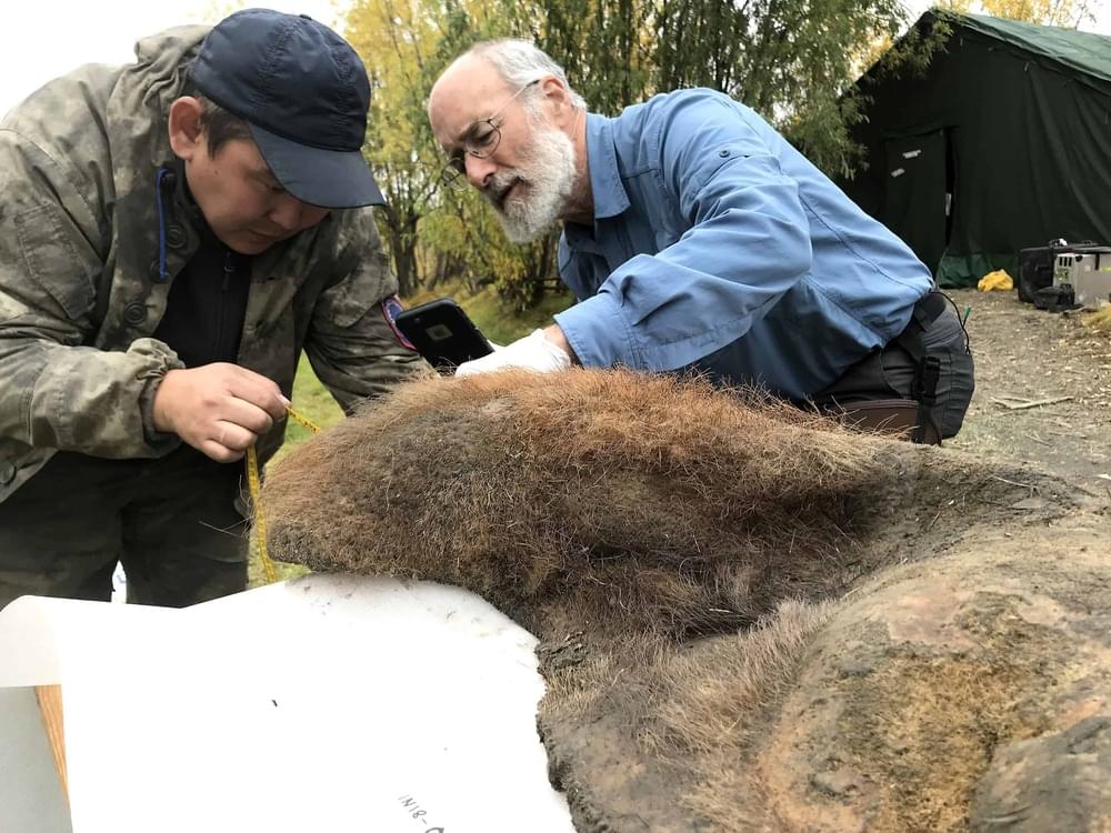
However, the 52,000-year-old skin sample at the heart of this research was taken from behind the ear of a mammoth discovered in Northern Siberia in 2018. An intensive analysis of the sample revealed the presence of complete fossil chromosomes. These chromosomes, each measuring billionths of a meter in length, had seemingly been frozen in a glass-like state for tens of thousands of years. Knowing the shape of an organism’s chromosomes can help researchers to assemble entire DNA sequences of extinct creatures, a task previously deemed nearly impossible due to DNA degradation over time.
“This is a new type of fossil, and its scale dwarfs that of individual ancient DNA fragments — a million times more sequence,” explained Erez Lieberman Aiden, a corresponding author on the study and director of the Center for Genome Architecture at the Baylor College of Medicine.