Toggle light / dark theme

Monocytes, a type of white blood cell, are alone capable of facilitating faster wound healing, says study.

Scientists from the University of Calgary, Canada, have discovered a promising new approach to treating bacterial skin infections. The research showed that monocytes alone are capable of facilitating faster wound healing. The researchers’ next step is to better understand how immune cells like neutrophils function during infection. Researchers have discovered a promising new approach to treating bacterial skin infections.

A team of scientists from the University of Calgary, Canada, revealed new insights which could lead to advancements in the treatment of bacterial infections and wounds, according to a study published in Nature science journal on Friday.

“It is exciting that we have made a fundamental discovery that could improve infections and tissue repair in humans, especially hard-to-treat cases,” said the study’s first author Dr. Rachel Kratofi in the press release. “Translating our research from bench to bedside will require many more experiments and involve a model more closely related to human disease.”

The research showed that monocytes alone are capable of facilitating faster wound healing. Monocytes contribute to wound healing by regulating leptin levels and blood vessel growth. They also produce ghrelin, a hormone that aids in wound healing. Historically, researchers believed that neutrophils and monocytes (white blood cells) were both recruited to clear bacteria from an infected site on the skin. When these cells work together, they serve as our bodies first line of defense against the immune system. The connection between metabolic hormones and tissue repair Ghrelin is produced by the stomach when you are hungry, whereas leptin is produced by fat cells after you eat a meal and feel full. This ghrelin-leptin balance has long been recognized as important for metabolism and diet. Still, its relationship to immune mechanisms and tissue repair has been unknown until now. Kratofil was able to visualize the immune response to Staphylococcus aureus (S. aureus) bacteria in an animal model using intravital microscopy, which allows observation of live cells and is a specialization of the university’s Kubes Lab.

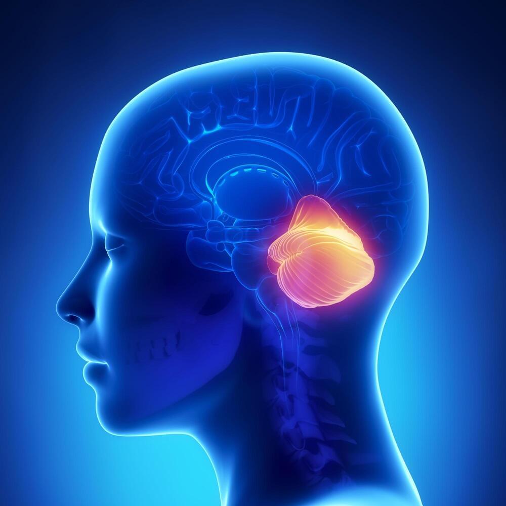
This article tells of possible way to increase brain intelligence through a certain mutation which in theory could be altered for biological singularity like effects in the future.


Humans carrying the CORD7 (cone-rod dystrophy 7) mutation possess increased verbal IQ and working memory. This autosomal dominant syndrome is caused by the single-amino acid R844H exchange (human numbering) located in the 310 helix of the C2A domain of RIMS1/RIM1 (Rab3-interacting molecule 1). RIM is an evolutionarily conserved multi-domain protein and essential component of presynaptic active zones, which is centrally involved in fast, Ca2+-triggered neurotransmitter release. How the CORD7 mutation affects synaptic function has remained unclear thus far. Here, we established Drosophila melanogaster as a disease model for clarifying the effects of the CORD7 mutation on RIM function and synaptic vesicle release.

To this end, using protein expression and X-ray crystallography, we solved the molecular structure of the Drosophila C2A domain at 1.92 Å resolution and by comparison to its mammalian homolog ascertained that the location of the CORD7 mutation is structurally conserved in fly RIM. Further, CRISPR/Cas9-assisted genomic engineering was employed for the generation of rim alleles encoding the R915H CORD7 exchange or R915E, R916E substitutions (fly numbering) to effect local charge reversal at the 310 helix. Through electrophysiological characterization by two-electrode voltage clamp and focal recordings we determined that the CORD7 mutation exerts a semi-dominant rather than a dominant effect on synaptic transmission resulting in faster, more efficient synaptic release and increased size of the readily releasable pool but decreased sensitivity for the fast calcium chelator BAPTA.

Circa 2015


“Their work has provided fundamental knowledge of how a living cell functions and is, for instance, used for the development of new cancer treatments,” the Royal Swedish Academy of Sciences said.

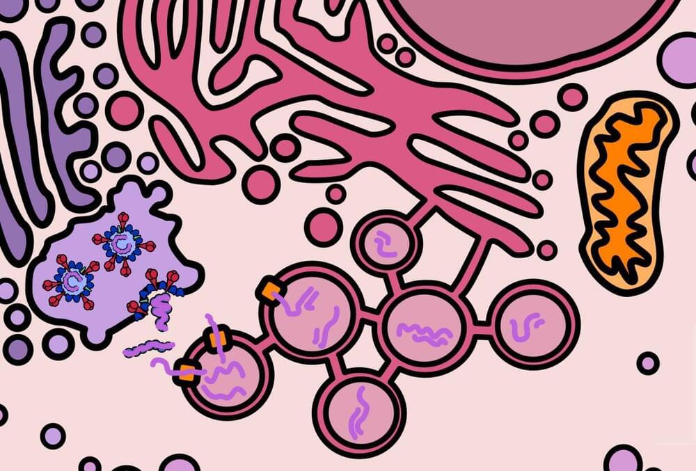
Thousands of alterations to a cell’s genome occur every day due to spontaneous changes and damage by radiation, free radicals and carcinogenic substances — yet DNA remains astonishingly intact.

To keep genetic materials from disintegrating, a range of molecular systems monitor and repair DNA, in processes that the three award-winning scientists helped map out.

This extensive research actually details the possibility of unknown origins of these elongated human skulls which many think to this day are some form of exterrestial in origin or at the very least unknown in origin which actually nearly uproots most know origin stories.


Modern European genetic structure demonstrates strong correlations with geography, while genetic analysis of prehistoric humans has indicated at least two major waves of immigration from outside the continent during periods of cultural change. However, population-level genome data that could shed light on the demographic processes occurring during the intervening periods have been absent. Therefore, we generated genomic data from 41 individuals dating mostly to the late 5th/early 6th century AD from present-day Bavaria in southern Germany, including 11 whole genomes (mean depth 5.56×). In addition we developed a capture array to sequence neutral regions spanning a total of 5 Mb and 486 functional polymorphic sites to high depth (mean 72×) in all individuals. Our data indicate that while men generally had ancestry that closely resembles modern northern and central Europeans, women exhibit a very high genetic heterogeneity; this includes signals of genetic ancestry ranging from western Europe to East Asia. Particularly striking are women with artificial skull deformations; the analysis of their collective genetic ancestry suggests an origin in southeastern Europe. In addition, functional variants indicate that they also differed in visible characteristics. This example of female-biased migration indicates that complex demographic processes during the Early Medieval period may have contributed in an unexpected way to shape the modern European genetic landscape. Examination of the panel of functional loci also revealed that many alleles associated with recent positive selection were already at modern-like frequencies in European populations ∼1,500 years ago.

Realistic and complex models of brain cells, developed at Cedars-Sinai with support from our scientists and our #openscience data, could help answer questions a… See more.


Cedars-Sinai investigators have created bio-realistic and complex computer models of individual brain cells—in unparalleled quantity.

Their research, published today in the peer-reviewed journal Cell Reports, details how these models could one day answer questions about neurological disorders—and even human intellect—that aren’t possible to explore through biological experiments.

“These models capture the shape, timing and speed of the electrical signals that neurons fire in order to communicate with each other, which is considered the basis of brain function,” said Costas Anastassiou, PhD, a research scientist in the Department of Neurosurgery at Cedars-Sinai, and senior author of the study. “This lets us replicate brain activity at the single-cell level.”

Around the same time, neuroscientists developed the first computational models of the primate visual system, using neural networks like AlexNet and its successors. The union looked promising: When monkeys and artificial neural nets were shown the same images, for example, the activity of the real neurons and the artificial neurons showed an intriguing correspondence. Artificial models of hearing and odor detection followed.

But as the field progressed, researchers realized the limitations of supervised training. For instance, in 2017, Leon Gatys, a computer scientist then at the University of Tübingen in Germany, and his colleagues took an image of a Ford Model T, then overlaid a leopard skin pattern across the photo, generating a bizarre but easily recognizable image. A leading artificial neural network correctly classified the original image as a Model T, but considered the modified image a leopard. It had fixated on the texture and had no understanding of the shape of a car (or a leopard, for that matter).

Self-supervised learning strategies are designed to avoid such problems. In this approach, humans don’t label the data. Rather, “the labels come from the data itself,” said Friedemann Zenke, a computational neuroscientist at the Friedrich Miescher Institute for Biomedical Research in Basel, Switzerland. Self-supervised algorithms essentially create gaps in the data and ask the neural network to fill in the blanks. In a so-called large language model, for instance, the training algorithm will show the neural network the first few words of a sentence and ask it to predict the next word. When trained with a massive corpus of text gleaned from the internet, the model appears to learn the syntactic structure of the language, demonstrating impressive linguistic ability — all without external labels or supervision.

Vanderbilt researchers have developed an active machine learning approach to predict the effects of tumor variants of unknown significance, or VUS, on sensitivity to chemotherapy. VUS, mutated bits of DNA with unknown impacts on cancer risk, are constantly being identified. The growing number of rare VUS makes it imperative for scientists to analyze them and determine the kind of cancer risk they impart.

Traditional prediction methods display limited power and accuracy for rare VUS. Even machine learning, an artificial intelligence tool that leverages data to “learn” and boost performance, falls short when classifying some VUS. Recent work by the lab of Walter Chazin, Chancellor’s Chair in Medicine and professor of biochemistry and chemistry, led by co-first authors and postdoctoral fellows Alexandra Blee and Bian Li, featured an active machine learning technique.

Active machine learning relies on training an algorithm with existing data, as with machine learning, and feeding it new information between rounds of training. Chazin and his lab identified VUS for which predictions were least certain, performed biochemical experiments on those VUS and incorporated the resulting data into subsequent rounds of algorithm training. This allowed the model to continuously improve its VUS classification.

Thor Balkhed/Linköping University.

Made of collagen protein from pig’s skin, the implant resembles the human cornea and is more than a pipe dream for an estimated number of 12.7 million people around the world who are blind due to their diseased corneas. The implant is a promising alternative to the transplantation of donated human corneas, which are scarce in under-developed and developing countries, where the need for them is greatest.