Sleep, fasting, exercise, green porridge, black coffee, a healthy social life—there is an abundance of advice out there on how to live a good, long life. Researchers are working hard to determine why some people live longer than others, and how we get the most out of our increasingly long lives.
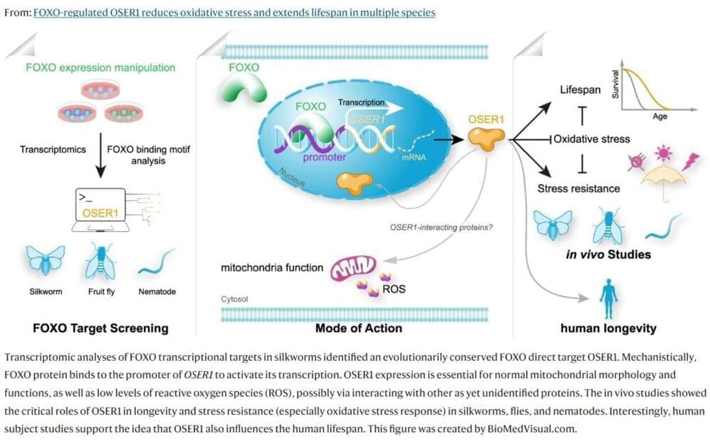
Now researchers from the Center for Healthy Aging, Department of Cellular and Molecular Medicine at the University of Copenhagen have discovered that a particular protein known as OSER1 has a great influence on longevity. The research is published in the journal Nature Communications.
“We identified this protein that can extend longevity. It is a novel pro-longevity factor, and it is a protein that exists in various animals, such as fruit flies, nematodes, silkworms, and in humans,” says Professor Lene Juel Rasmussen, senior author behind the new study.