Toggle light / dark theme

Engineers have created intelligent 3D printers that can quickly detect and correct errors, even in previously unseen designs, or unfamiliar materials like ketchup and mayonnaise, by learning from the experiences of other machines.

The engineers, from the University of Cambridge, developed a machine learning algorithm that can detect and correct a wide variety of different errors in real time, and can be easily added to new or existing machines to enhance their capabilities. 3D printers using the algorithm could also learn how to print new materials by themselves. Details of their low-cost approach are reported in the journal Nature Communications.

3D has the potential to revolutionize the production of complex and customized parts, such as aircraft components, personalized medical implants, or even intricate sweets, and could also transform manufacturing supply chains. However, it is also vulnerable to production errors, from small-scale inaccuracies and mechanical weaknesses through to total build failures.

Down the line, the researchers hope to develop an injectable protein solution that forms a gel inside the body. The ability to design hydrogels with specific functions opens up for a range of possible applications. Such a gel could, for example, be used to achieve a controlled release of drugs into the body. In the chemical industry, it could be fused to enzymes, a form of proteins used to speed up various chemical processes.

“In the slightly longer term, I think injectable gels can become very useful in regenerative medicine,” says the study’s first author Tina Arndt, a PhD student in Anna Rising’s research group at Karolinska Institute. “We have a long way to go, but the fact that the protein solution quickly forms a gel at body temperature and that the spider silk has been shown to be well tolerated by the body is promising.”

The ability of spiders to spin incredibly strong fibers from a silk protein solution in fractions of a second has sparked an interest in the underlying molecular mechanisms. The researchers at KI and SLU have been particularly interested in the spiders’ ability to keep proteins soluble so that they do not clump together before the spinning of the spider silk. They have previously developed a method for the production of valuable proteins which mimics the process the spider uses to produce and store its silk proteins.

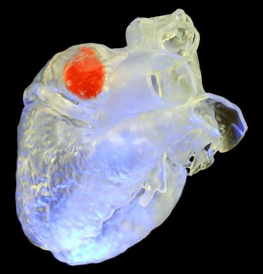
Although chemotherapy can be a lifesaving treatment for patients with cancer, some of these medications can damage the heart. A team led by researchers at Massachusetts General Hospital (MGH) recently developed a nanoparticle probe that can detect an indicator of heart damage from chemotherapy.

Experiments with the probe also revealed that in mice with cancer, intermittent fasting before chemotherapy can prevent this damage indicator from arising, leading to preserved cardiac function and prolonged survival.

The study, which is published in Nature Biomedical Engineering, focused on autophagy—a process that cells use to remove unnecessary or dysfunctional components. A delicate balance exists between the protective and deleterious effects of this process: reduced levels of autophagy have been implicated in and other conditions; however, autophagy can also be a primary mechanism of cell death.

A simple statistical test shows that contrary to current practice, the “gaps” within DNA protein and sequence alignments commonly used in evolutionary biology can provide important information about nucleotide and amino acid substitutions over time. The finding could be particularly relevant to those studying distantly related species. The work appears in Proceedings of the National Academy of Sciences.

Biologists studying evolution do so by looking at how DNA and protein sequences change over time. These changes can be sequence length changes—when specific are deleted or added at certain positions—or substitutions, where one nucleotide type is exchanged for a different type at a given point.

“Think of the DNA sequence and its evolution as a sentence being copied by different people over time,” says Jeff Thorne, professor of biological sciences and statistics at NC State and a co-corresponding author of the research. “Over time, a letter in a word will change—that’s a substitution. Leaving out or adding letters or words correspond to deletions or insertions.”

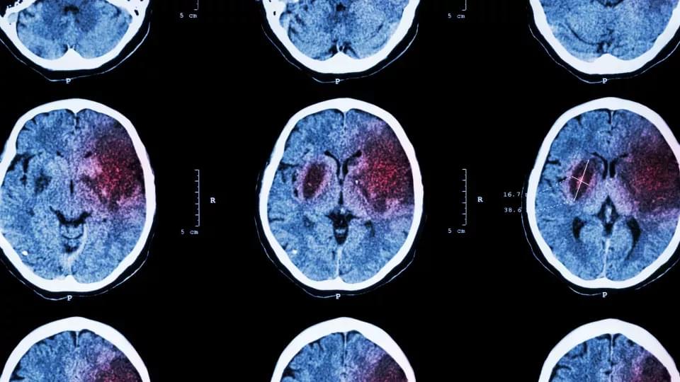
An artificial intelligence (AI) algorithm to detect subtle brain abnormalities that cause epileptic seizures has been developed. The abnormalities, known as focal cortical dysplasias (FCDs), can often be treated with surgery but are difficult to visualize on an MRI. The new algorithm is expected to give physicians greater confidence in identifying FCDs in patients with epilepsy.

The work, which was part of the Multicentre Epilepsy Lesion Detection (MELD) project, appeared in Brain Interpretable surface-based detection of focal cortical dysplasias: a Multi-centre Epilepsy Lesion Detection study.” Konrad Wagstyl, PhD, and Sophie Adler, PhD, both from University College London, led an international team of researchers on the work.

To develop the algorithm, the team quantified features of the brain cortex—such as thickness and folding—in more than 1,000 patient MRI scans from 22 epilepsy centers around the world. They then trained the algorithm on examples labeled by expert radiologists as either being healthy or having FCD.

To evaluate the accuracy of the models28, we sampled from the BNN or deep ensemble to determine their uncertainty predictions (Extended Data Fig. 3a, b). Correct predictions are oriented toward the lower and higher range of the output, representing greater certainty about samples’ states, whereas incorrect predictions tend towards the 0.5 threshold. We can therefore assume higher confidence in a model’s predictions by removing the predictions in the middle using thresholds. We evaluated a range of thresholds with several models (Extended Data Fig. 3c–f), which show a substantial increase in accuracy due to the ambiguous samples being discarded, including the ensemble of normalized models reaching accuracy of 97.2%. A similar approach was applied to other models, including the IR and RS models (Extended Data Fig. 3g, h), raising accuracy by 10–15%, although this reduces the number of cells considered.

To better understand the development of the senescent phenotype and how nuclear morphology changes over time, we analyzed human fibroblasts induced to senescence by 10 Gy IR and imaged at days 10, 17, 24 and 31. The predictor identifies senescence at all four times points with probability that increases from days 10 to 17 but declines by day 31 (Extended Data Fig. 4a). Interestingly, examining the probability distribution of the predictor it was apparent that a growing peak of nonsenescent cells appear after day 17, suggesting that a small number of cells were able to escape senescence induction and eventually overgrow the senescent cells (Extended Data Fig. 4b). Indeed, when investigating markers of proliferation, we see that over the time course, PCNA declines until day 17, after which the expression starts to return (Extended Data Fig. 4c). p21Cip1 follows an inverse pattern with stain intensity increasing initially and then declining slightly by day 31 (Extended Data Fig. 4D). We also saw a decrease in DAPI intensity for days 10 and 17, indicating senescence, but a reversion to control level by day 31 (Extended Data Fig. 4e). To confirm that the predictor accurately determined senescence even 31 days after IR, we evaluated if markers of proliferation and senescence correlated with predicted senescence. Accordingly, cells with predicted senescence had higher p21Cip1 levels, lower PCNA and lower DAPI intensities and vice versa (Extended Data Fig. 4f–h). Morphologically, area and aspect are higher for predicted senescence, whereas convexity is lower (Extended Data Fig. 4i–k). Finally, a simple nuclei count confirms growth, following IR treatment (Extended Data Fig. 4l). Overall, the senescence predictor captures the state during development in agreement with multiple markers and morphological signs.

Senescent cells are associated with the appearance of persistent nuclear foci of the DNA damage markers γH2AX and 53BP1 (refs. 31,32). Our base data set including control, RS and IR lines were examined for damage foci using high-content microscopy, where we found the mean count for controls to be below 1 for each marker, whereas RS had 4.0 γH2AX and 2.0 53BP1 foci and IR had 3.4 γH2AX and 3.0 53BP1 foci (Fig. 4a, b and Extended Data Fig. 5a). We calculated the Pearson correlation between predicted senescence and γH2AX and 53BP1 foci counts and found that across all conditions, there is a moderately strong correlation of around 0.5 (Fig. 4c). This association is also visible when simply plotting foci counts and senescence prediction, which shows predicted senescence flipping from low to high, along with shifts in foci counts (Extended Data Fig. 5b). Our feature reduction masked internal nuclear structure, but it is nonetheless notable that senescence prediction correlates with foci count. We also compared the correlation between predicted senescence and area, where we see a correlation of around 0.5. In sum, there is a considerable correlation between foci counts and senescence.

In this review, we undertake a critical appraisal of eight published studies providing first evidence that a history of attention-deficit/hyperactivity disorder (ADHD) may increase risk for the later-life development of a neurodegenerative disease, in particular Lewy body diseases (LBD), by up to five-fold. Most of these studies have used data linked to health records in large population registers and include impressive sample sizes and adequate follow-up periods. We identify a number of methodological limitations as well, including potential diagnostic inaccuracies arising from the use of electronic health records, biases in the measurement of ADHD status and symptoms, and concerns surrounding the representativeness of ADHD and LBD cohorts. Consequently, previously reported risk associations may have been underestimated due to the high likelihood of potentially missed ADHD cases in groups used as “controls”, or alternatively previous estimates may be inflated due to the inclusion of confounding comorbidities or non-ADHD cases within “exposed” groups that may have better accounted for dementia risk. Prospective longitudinal studies involving well-characterized cases and controls are recommended to provide some reassurance about the validity of neurodegenerative risk estimates in ADHD.

Attention-deficit hyperactivity disorder (ADHD) is a psychiatric disorder beginning in childhood that is characterized by core symptoms of inattention, impulsivity, and hyperactivity (Biederman and Faraone, 2005; American Psychiatric Association, 2013; Faraone et al., 2015). Diagnostic criteria require symptoms to present in early childhood, before age 12, and cause impairment in daily activities in more than one setting (e.g., home, school, social environment, and/or interpersonal relationships; American Psychiatric Association, 2013). Although it is largely considered a childhood disorder, 40–60% of cases of ADHD persist into adulthood (Culpepper and Mattingly, 2010; Michielsen et al., 2012; Volkow and Swanson, 2013; Asherson et al., 2016), and the overall prevalence of adult ADHD ranges from 2 to 4% (Kieling and Rohde, 2012; Fayyad et al., 2017).

ADHD may persist into later life as well. Roughly 3% of adults over age 50 suffer from significant symptoms of attention-deficit/hyperactivity disorder (ADHD; Michielsen et al., 2012; Kooij et al., 2016), often presenting as executive dysfunction (e.g., absent-mindedness) and memory impairments (e.g., forgetfulness or difficulty learning new things; Rosler et al., 2010; Thorell et al., 2017; Callahan et al., 2021). These symptoms overlap with those of early neurodegenerative disease (Ivanchak et al., 2012; Pollack, 2012; Goodman et al., 2016; Callahan et al., 2017), and it is currently unclear whether ADHD is associated with an increased neurodegenerative risk, or if it is being misdiagnosed due to symptom overlap (Callahan et al., 2017). Clarifying this issue is crucial to reduce dementia misdiagnoses, and to guide treatment, which will differ depending on whether the disease course is assumed to be neurodegenerative or not.

AgeX Therapeutics, a preclinical biotech looking to turn back the clock on aging, may have to wind down, announcing “substantial doubt” about its ability to continue as money runs dry and debts mount.

The California biotech made $12,000 in revenue for the second quarter and recorded $1.6 million in operating expenses over the same period, according to financial results posted August 12.

During the first quarter, the biotech borrowed the final half million of credit available under a 2020 agreement with Juvenescence—a separate anti-aging biotech—and entered a new deal in which Juvenescence will provide $13.2 million in credit for a year. AgeX drew an initial $8.2 million of the line of credit and used $7.2 million to refinance the principal and the loan origination fees under a 2019 loan agreement with Juvenescence.

The seven Expedition 67 residents kicked off a busy week of critical research benefitting humans living on and off the Earth. The orbital residents also continued supporting the International Space Station’s vast array of flight, research, and life support systems.

Astronauts Bob Hines of NASA and Samantha Cristoforetti of ESA (European Space Agency) took turns on Monday cleaning hardware and supporting samples for a biology study that is exploring skin healing in space. Observations may provide insights improving wound healing therapies for astronauts and Earthlings. Hines then spent the afternoon installing seed cartridges and root modules for the XROOTS space agriculture investigation to begin a 30-day growth period of radishes and mizuna greens. The research uses hydroponics and aeroponics techniques to learn how to produce crops on a larger scale on future missions to the Moon, Mars, and beyond.

NASA Flight Engineer Kjell Lindgren opened up the Kibo laboratory module’s airlock and retrieved an external science platform and installed a small satellite deployer on the research gear. The deployer will be placed outside Kibo in the vacuum of space before deploying a set of CubeSats into low-Earth orbit for a variety of research and education programs.