🧠 Neuromodulation through the eyes 👀
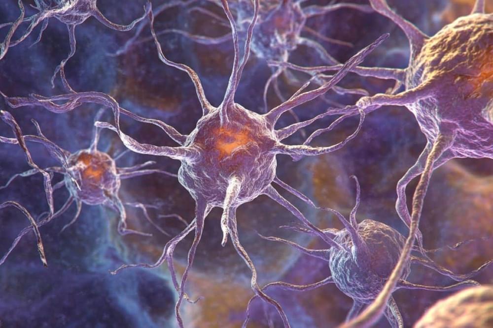
Neuroplasticity, also known as neural plasticity or brain plasticity, is a process that involves adaptive structural and functional changes to the brain.
Founded and directed by Deborah Zelinsky, O.D., F.N.O.R.A., F.C.O.V.D.
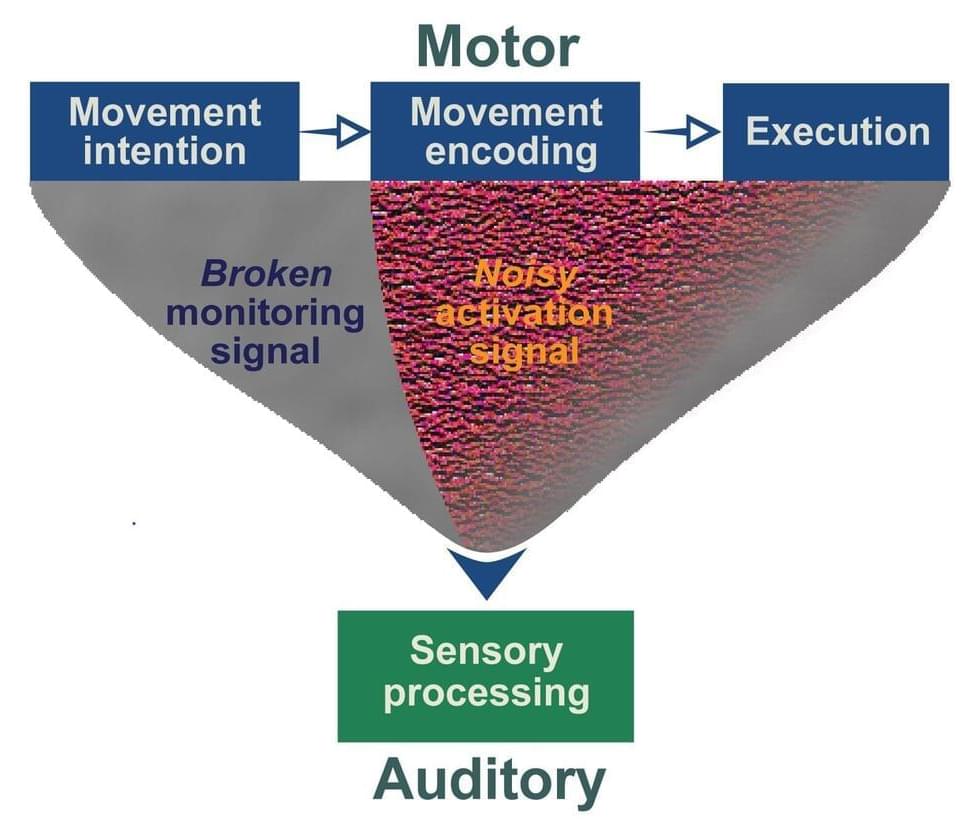
Just as with eye-hand coordination, integration of vision and sound – eye-ear connection – must be developed. If the two senses are out of sync, a person can experience difficulties in academics, social situations and activities such as sports.
Balance between vision and hearing is necessary for a person to learn letter sounds, for example, while applying those sounds to the words they see on a page. In social situations, a person can better understand what another is saying – and meaning — by watching body language and facial expression. Autistic patients cannot discern the nuances of a joke because they simply listen. They do not connect sound and vision, because the environment around them is too confusing.
A student whose eyesight is more sensitive than his or her hearing may be easily distracted by activities and moving objects in the environment and unable to concentrate on what the teacher is saying. People whose peripheral vision is not sufficiently “tuned in” may have to turn their head before finding what is causing a certain sound.