Toggle light / dark theme

Tissue engineers create artificial organs and tissues that can be used to develop and test new drugs, repair damaged tissue and even replace entire organs in the human body. However, current fabrication methods limit their ability to produce free-form shapes and achieve high cell viability.

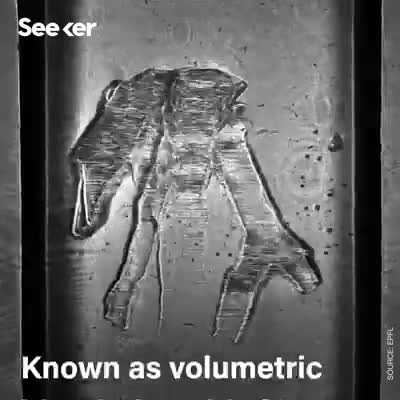
Researchers at the Laboratory of Applied Photonics Devices (LAPD), in EPFL’s School of Engineering, working with colleagues from Utrecht University, have come up with an that takes just a few seconds to sculpt complex shapes in a biocompatible hydrogel containing stem . The resulting tissue can then be vascularized by adding endothelial cells.

The team describes this high-resolution printing method in an article appearing in Advanced Materials. The technique will change the way cellular engineering specialists work, allowing them to create a new breed of personalized, functional bioprinted organs.

While in its early stages, bioprinting of human tissue is an emerging technology that is opening up some exciting possibilities, including the potential to one day 3D print entire human organs. This scientific objective has now grown a little bit closer, with researchers at Carnegie Mellon University reporting a breakthrough that enabled the printing of full-scale heart components that in some cases functioned similarly to the real thing.

Over 4000 patients in the United States alone are waiting for a heart transplant, while millions of others worldwide need hearts but are ineligible for the waitlist. The need for replacement organs is immense, and new approaches are needed to engineer artificial organs that are capable of repairing, supplementing, or replacing long-term organ function.


A team of researchers from Carnegie Mellon University has published a paper in Science that details a new technique allowing anyone to 3D bioprint tissue scaffolds out of collagen, the major structural protein in the human body. This first-of-its-kind method brings the field of tissue engineering one step closer to being able to 3D print a full-sized, adult human heart.

The technique, known as Freeform Reversible Embedding of Suspended Hydrogels (FRESH), has allowed the researchers to overcome many challenges associated with existing 3D bioprinting methods, and to achieve unprecedented resolution and fidelity using soft and living materials.

Each of the organs in the , such as the heart, is built from specialized cells that are held together by a biological scaffold called the extracellular matrix (ECM). This network of ECM proteins provides the structure and biochemical signals that cells need to carry out their normal function. However, until now it has not been possible to rebuild this complex ECM architecture using traditional biofabrication methods.

Kyle Reese: The Terminator’s an infiltration unit, part man, part machine. Underneath, it’s a hyperalloy combat chassis — micro processor-controlled, fully armored. Very tough. But outside, it’s living human tissue — flesh, skin, hair, blood, grown for the cyborgs…


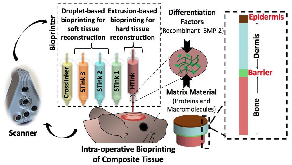
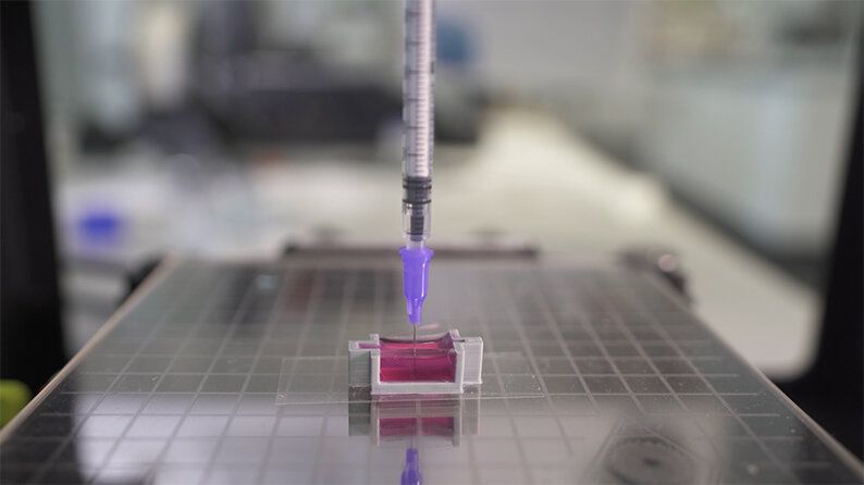
3D bioprinting is the automated fabrication of multicellular tissue via spatially defined deposition of cells. The ability to spatially control deposition in the x, y and z axes allows for creation of tissue-specific patterns or compartments, with in vivo-like architecture that mimics key aspects of native biology.

3D bioprinted tissues exhibit a microenvironment more suited to in vivo-like cellular function in comparison to traditional 2D monoculture (or monolayer co-cultures), as well as maintenance of a more defined architecture than is observed in self-aggregated co-culture models.

Read more