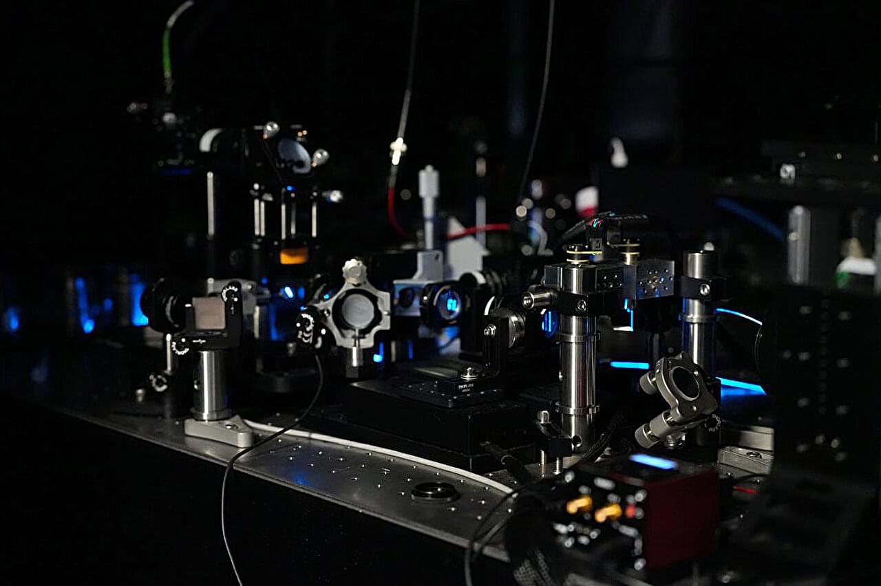
An international team of researchers led by UT Southwestern Medical Center has developed a versatile light-sheet microscope that captures 3D views of biological samples, from activity inside living cells to larger tissues. Designed to make detailed imaging much simpler and faster, the approach could help laboratories stretch equipment budgets and expand research that may ultimately inform patient care.
“Our work makes a popular microscope technique, called light-sheet fluorescence microscopy, more versatile and user-friendly,” said corresponding author Reto Fiolka, Ph.D., associate professor in the Lyda Hill Department of Bioinformatics and of Cell Biology at UT Southwestern. “It extends what a single instrument can do in terms of sample coverage and imaging applications.”
Described in Nature Methods, the multi-immersion oblique plane microscope (miOPM) addresses a long-standing challenge: Advanced microscopes are often designed for a narrow range of tasks. Studying different samples can require separate instruments, specialized preparation and considerable technical expertise.