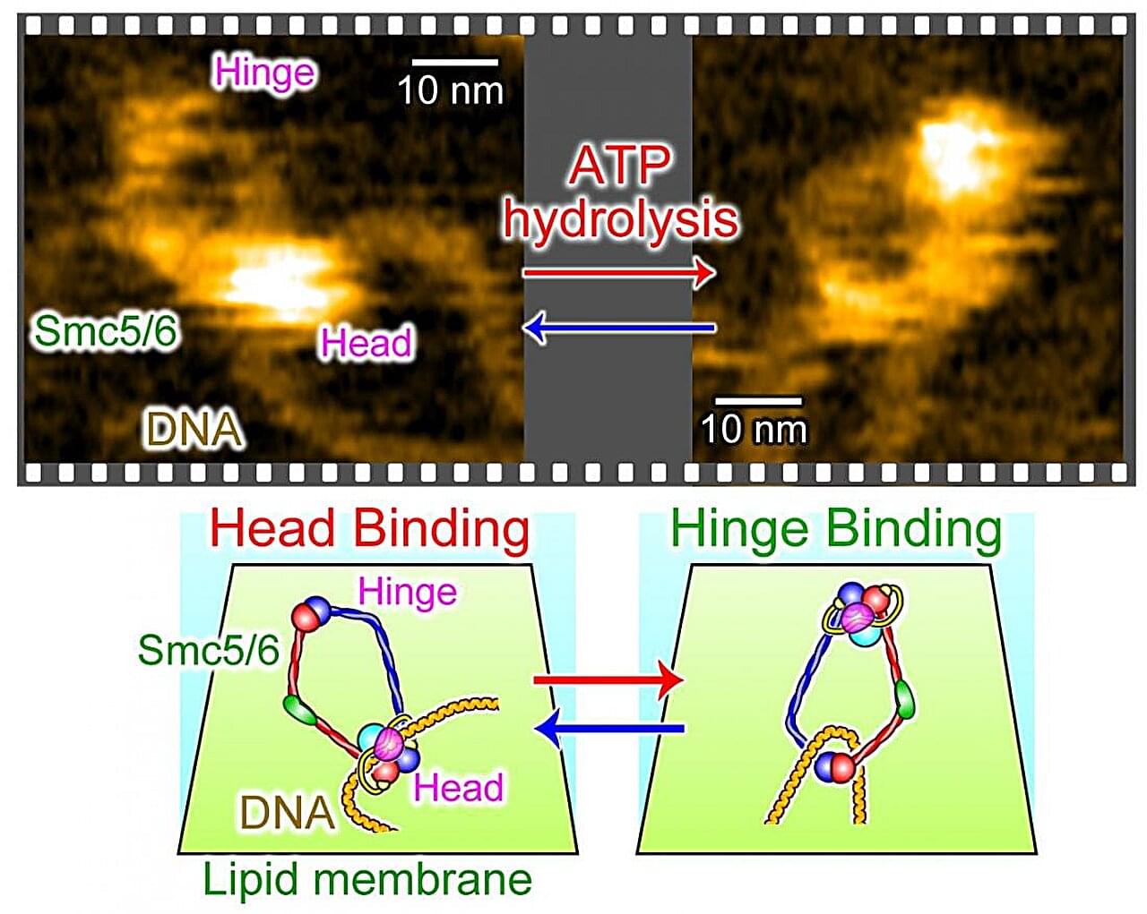
Researchers at Kanazawa University and the National Institute of Genetics in Japan have used high-speed atomic force microscopy to observe how a group of proteins known as Smc5/6 interacts with DNA. The images reveal how this structure opens and closes, captures DNA and brings separate sections together. The researchers also observed Smc5/6 holding twisted sections of DNA in place, producing more tightly packed arrangements. The findings, published in Nature Communications, provide new insights into the molecular processes that contribute to the maintenance and repair of genetic material.
Every living cell must protect and maintain its DNA. This genetic material contains the instructions needed for life, but it is constantly subjected to stresses that can damage its structure. DNA must also be copied accurately when cells divide. To accomplish these tasks, cells rely on proteins that interact with DNA, control its arrangement and support the processes needed to maintain its integrity.
One of these is Smc5/6, a structure made up of several proteins that work together. It belongs to a family of proteins involved in maintaining chromosomes, the structures that carry DNA inside cells. Although scientists have known that Smc5/6 is important for DNA maintenance and repair, precisely how it captures and handles DNA has remained difficult to observe.