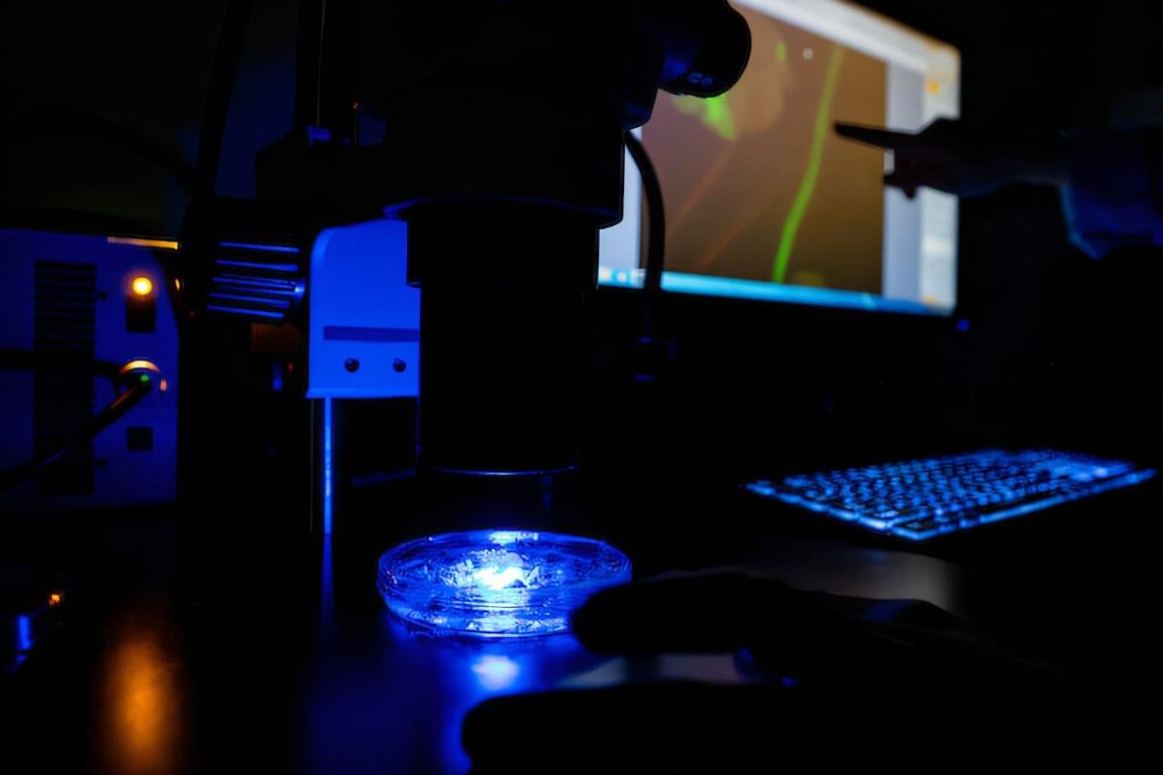
Researchers at Texas A&M University have cracked a problem that has limited biological imaging for years: seeing fine cellular detail while looking deep inside living tissue. The new microscopy technique resolves structures smaller than 100 nanometers, roughly a thousand times thinner than a human hair, without giving up the ability to image deep into intact samples.
The team, led by Aart Verhoef, Ph.D., assistant professor of biophotonics in the Department of Soil and Crop Sciences in the Texas A&M College of Agriculture and Life Sciences and joint professor in the Department of Physics and Astronomy, published the findings in the Journal of Biomedical Optics.
Their method, called 2PE-ISM-SOFI, short for two-photon excited image scanning microscopy combined with super-resolution optical fluctuation imaging, layers two established techniques onto a standard two-photon microscope to sharpen its resolution without shrinking its reach.