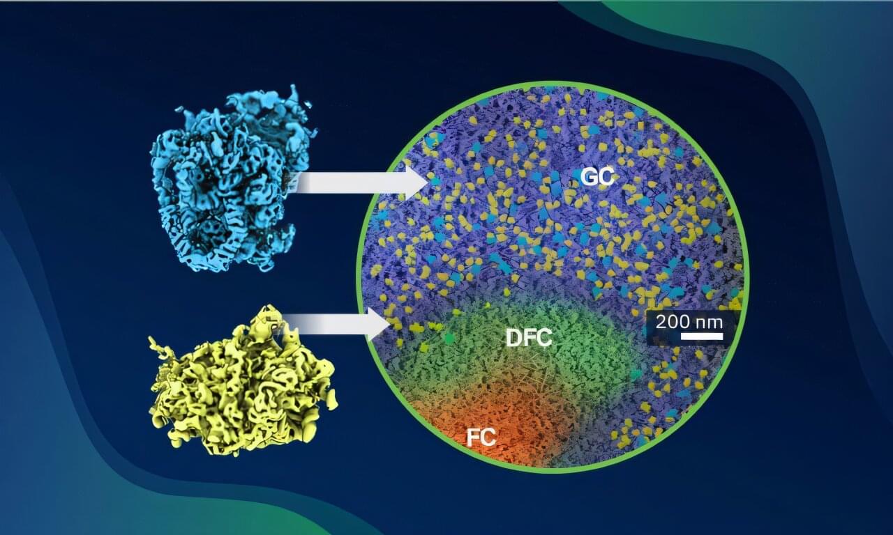
For decades, ribosomes have been under the lens of many scientists. Ribosomes are highly sophisticated and complex molecular machines that allow cells to synthesize proteins. However, putting ribosomes together requires intricate assembly processes. While many underlying mechanisms for building ribosomes have been discovered ex situ (outside their native cellular context), our understanding of how ribosomes are assembled in situ has so far remained limited.
Through a collaboration between the research groups of Christoph Müller, Sara Cuylen-Häring and Julia Mahamid at EMBL Heidelberg, researchers have now pushed these limits. By combining cryo-electron tomography (cryo-ET) with correlative light and electron microscopy (CLEM), they were able to image the nucleolus—the site of ribosome assembly inside cells—at sufficiently high resolution to visualize the ribosome assembly pathway inside intact human cells for the first time.
The results—a collection of molecular snapshots—yielded subnanometer-resolution in-cell structures of tens of intermediates along the ribosome assembly pathways, complementing and expanding previous insights into ribosome assembly obtained from ex situ structures. The new results have now been published in the journal Science.