To delineate HDAC10’s function in T cells, we engineered CD4+ T cell-specific Hdac10 knockout (KO) mice (Hdac10fl/fl-CD4Cre) by crossing Hdac10fl/fl mice with CD4Cre mice (Figure S2 A), which were genotyped using PCR (Figure S2 B). The deletion of HDAC10 in CD4+ T cells was confirmed by western blotting (WB) and RT-qPCR (Figures S2 C and S2D). We observed that Hdac10 absence in CD4+ T cells had no effects on T cell development and differentiation in thymus (Figure S2 E and S2F) and T cells composition, apoptosis, or proliferation in spleen and mesenteric lymph nodes (mLN) (Figures S2 G–S2R) at steady state. Collectively, these data demonstrate that HDAC10 is dispensable for steady-state T cell development.
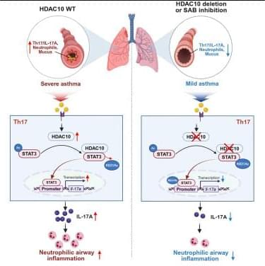
To determine HDAC10’s role in CD4+ T cells during allergic airway inflammation, we employed a neutrophilic asthma mouse model. Hdac10fl/fl-CD4Cre asthmatic mice exhibited lowered airway resistance compared with Hdac10fl/fl asthmatic mice (Figure 2A). Total cell counts and the number of neutrophils in BALF were profoundly reduced in Hdac10fl/fl-CD4Cre asthmatic mice compared with the littermates (Figures 2B and 2C). Hematoxylin and eosin (HE) staining confirmed attenuated inflammation score in Hdac10fl/fl-CD4Cre asthmatic mice (Figures 2D and 2E). Peribronchial trichrome (Masson) and periodic acid Schiff (PAS) staining showed a reduction in collagen deposition and mucus production in Hdac10fl/fl-CD4Cre asthmatic mice (Figures 2F–2I). Furthermore, IHC for myeloperoxidase (MPO) showed reduced neutrophil infiltration in lung tissues of Hdac10fl/fl-CD4Cre asthmatic mice (Figures 2J and 2K).