Researchers have developed a compact, handheld mid-infrared imaging spectrometer that can produce high-resolution chemical maps of a sample without using stains or labels. With further development, the handheld device might provide a portable and easy-to-use way to map the molecular makeup of tissues and other samples.
“Ultimately, this technology could make it possible to assess tissue during cancer surgery,” said research team leader Rohith Reddy of the University of Houston. “After removing a suspected tumor, a surgeon could scan the freshly excised tissue to help determine whether it is malignant or whether cancer cells remain at the surgical margin. This complementary information would be available while the patient is still in the operating room instead of having to wait for results from laboratory testing.”
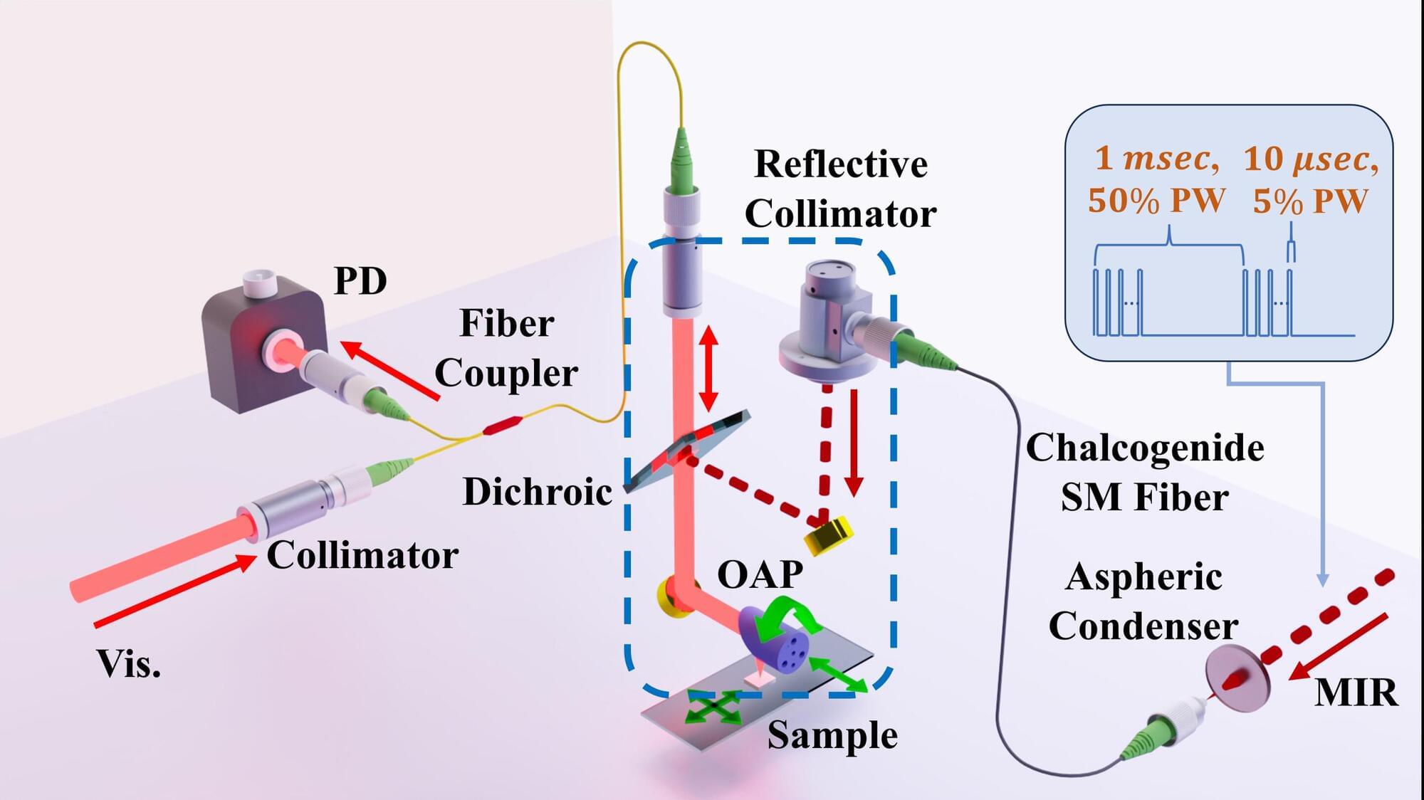
In Optica, the researchers describe how they transformed a photothermal mid-infrared spectroscopic imaging (MIRSI) system, normally a benchtop instrument occupying more than 9 square feet (0.8 square meters), into a handheld probe measuring 8 by 8 inches (20 by 20 centimeters). The probe holds the full optical head and connects by a flexible fiber tether to a compact base unit housing the lasers and control electronics. In side-by-side tests, it delivered image quality and chemical detail comparable to a state-of-the-art benchtop MIRSI system.