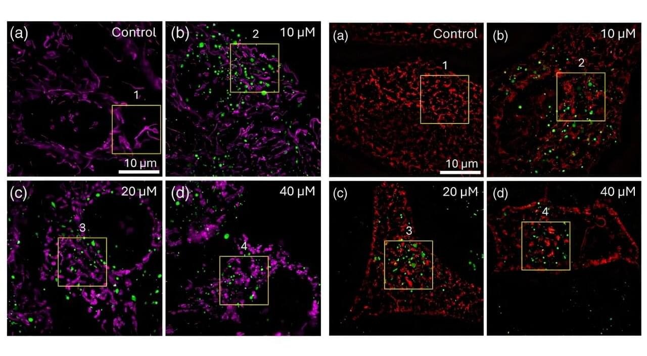
Cancer drugs are often designed to block specific molecular targets, but what happens after they enter a cell is not always well understood. A new study published in Biophotonics Discovery demonstrates how advanced imaging technology can help answer that question.
Using super-resolution microscopy, researchers tracked the cancer drug sunitinib inside living cells and observed how it altered several of the cell’s most important structures. The work provides a detailed view of drug behavior at the microscopic level and highlights the growing role of optical imaging in drug development and cellular engineering.
Sunitinib is used to treat several cancers, including kidney cancer. Its therapeutic activity has traditionally been linked to its ability to inhibit enzymes that drive tumor growth. However, researchers have increasingly recognized that where a drug travels inside a cell can also influence its effectiveness and side effects.