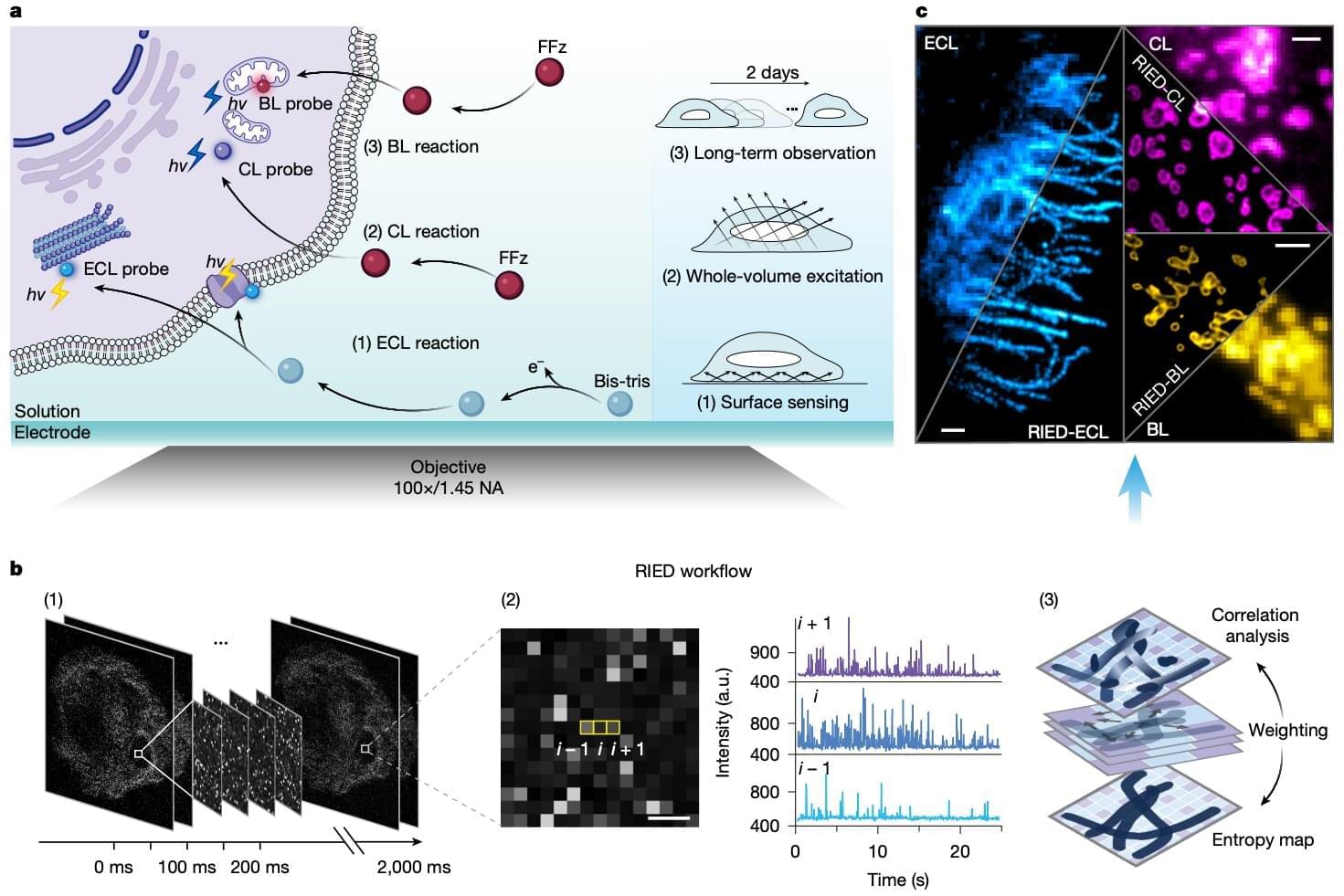
We reach for brighter, better lighting for sharper pictures, whether we’re photographing a puppy or a microscopic cell. In most cases, the light comes from outside the object being photographed. A recent study explored a different approach, using the cells’ own chemical glow to illuminate their internal structures. This new experimental framework, called REID, lets microscopes capture super-sharp, high-resolution details of cell structures by bypassing the need for harsh external lasers that can sometimes damage the cells being imaged.
Chemical reactions inside cells are normally very dim, but researchers discovered that swapping a common chemical co-reactant for a biological buffer called Bis-Tris boosted the cellular glow by 1,000 times. This let them capture sharp, unblurred images in just 20 milliseconds. The REID framework combined computer algorithms with electricity-triggered, chemical and biological (BL) luminescence to sharpen images down to 100 nanometers.
The team tested REID’s sensitivity, or how little of a biomarker it could still detect, using the cancer marker CEA, and compared it with standard fluorescence microscopy. They found that although the REID setup was less efficient at producing light, it was eight times more sensitive at detecting the cancer marker than the standard technique. The findings are published in Nature.