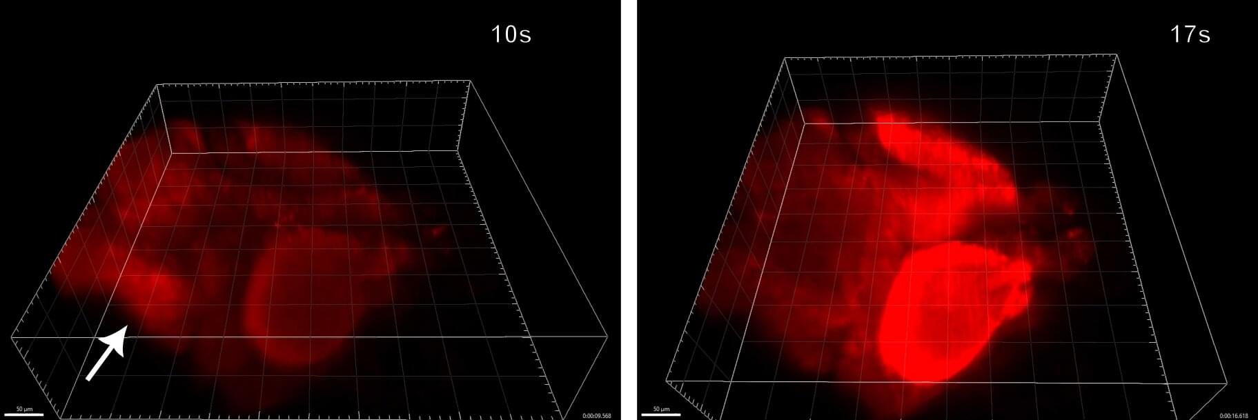
Seizures can race through the brain in seconds, making them difficult to capture in detail. To overcome this challenge, researchers have developed a new high-resolution light-sheet imaging system that is fast enough to image seizure propagation in the brain of a larval zebrafish in 3D.
“We developed a light-sheet microscope that allows rapid volumetric imaging with real-time correction of aberrations—imperfections in the way a microscope forms an image,” said research team leader Peter Kner from the University of Georgia. “Most imaging of seizure events in zebrafish has only captured 2D images, but our system allows 3D high-resolution imaging over a larger volume than was previously possible.”
In their article published in the journal Biomedical Optics Express, the researchers show that their new microscopy system can capture volumes up to 499 × 499 × 150 microns3 at a rate of four volumes per second with near-diffraction-limited resolution. They used the system to observe how seizures spread through the nervous system in zebrafish larvae, which are commonly used in neuroscience research.