Imagine if meaning — the elusive essence of language and thought — could be broken down into mathematical building blocks as fundamental as prime numbers. What if computers could “reason” by synchronizing oscillators, much like neurons firing in harmony in our brains?
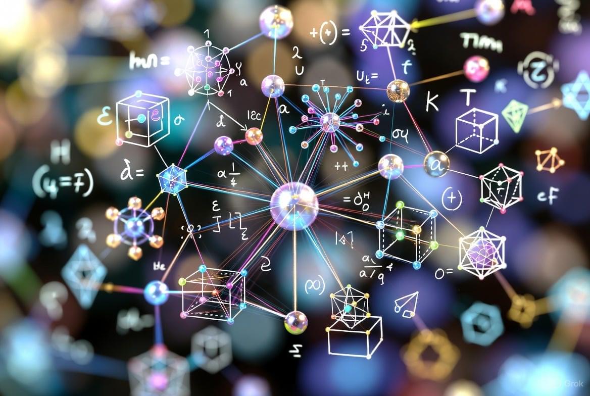
That’s the bold idea behind TinyAleph, a new framework and library I’ve developed for semantic computing. Unlike today’s AI models that gobble up massive datasets to mimic understanding, TinyAleph grounds meaning in pure math: primes, hypercomplex algebra, and dynamic oscillators.
In this article, I’ll walk you through the core ideas of TinyAleph, stripping away the academic jargon to show why this could be a game-changer for AI, cryptography, and even quantum-inspired simulations. No PhD required — just an open mind.