One of the key predictions of general relativity, the bending of light around massive, compact objects, is observed for a supermassive black hole in the galaxy I Zwicky 1.


The Air Force Research Laboratory at Kirtland Air Force Base has released a new analysis of the Department of Defense’s investments into directed energy technologies, or DE. The report, titled “Directed Energy Futures 2060,” makes predictions about what the state of DE weapons and applications will be 40 years from now and offers a range of scenarios in which the United States might find itself either leading the field in DE or lagging behind peer-state adversaries. In examining the current state of the art of this relatively new class of weapons, the authors claim that the world has reached a “tipping point” in which directed energy is now critical to successful military operations.
One of the document’s most eyebrow-raising predictions is that a “force field” could be created by “a sufficiently large fleet or constellation of high-altitude DEW systems” that could provide a “missile defense umbrella, as part of a layered defense system, if such concepts prove affordable and necessary.” The report cites several existing examples of what it calls “force fields,” including the Active Denial System, or “pain ray,” as well as non-kinetic counter-drone systems, and potentially counter-missile systems, that use high-power microwaves to disable or destroy their targets. Most intriguingly, the press release claims that “the concept of a DE weapon creating a localized force field may be just on the horizon.”
The Air Force wants Hermeus Corporation to prove the concept for a high-speed transport, and maybe more.
The U.S. Air Force is looking to field a new type of low-cost yet advanced drone to be used as an “Off-Board Sensing Station,” or OBSS. Details remain very limited, and the few publicly available Air Force Research Laboratory documents on the program state that specifics are only available to approved contractors. Still, according to Kratos, one of the companies involved with the effort, the new unmanned platform could potentially end up being as revolutionary as the firm’s stealthy XQ-58 Valkyrie has been.
The remarks about the OBSS program were made by Eric DeMarco, President and Chief Executive Officer of Kratos Defense & Security Solutions, during a company earnings call this week. DeMarco says that if the program is successful, the company believes it “could ultimately be as significant and transformational to Kratos as we expect Valkyrie to be.” The CEO added that the OBSS program is a signal that “the total addressable market opportunity for Kratos’ class of tactical drones is rapidly expanding and clarifying, as the Department of Defense strives for affordable force multiplier systems and technologies.”


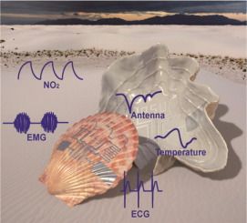
Deployment of functional circuits on a 3D freeform surface is of significant interest to wearable devices on curvilinear skin/tissue surfaces or smart Internet-of-Things with sensors on 3D objects. Here we present a new fabrication strategy that can directly print functional circuits either transient or long-lasting onto freeform surfaces by intense pulsed light-induced mass transfer of zinc nanoparticles (Zn NPs). The intense pulsed light can locally raise the temperature of Zn NPs to cause evaporation. Lamination of a kirigami-patterned soft semi-transparent polymer film with Zn NPs conforming to a 3D surface results in condensation of Zn NPs to form conductive yet degradable Zn patterns onto a 3D freeform surface for constructing transient electronics. Immersing the Zn patterns into a copper sulfate or silver nitrate solution can further convert the transient device to a long-lasting device with copper or silver. Functional circuits with integrated sensors and a wireless communication component on 3D glass beakers and seashells with complex surface geometries demonstrate the viability of this manufacturing strategy.

Skydweller Aero’s latest flight test of a modified solar-powered aircraft will provide the real-world data necessary for the U.S.-Spanish startup’s engineers to start developing and testing their proprietary autonomous flight software.
Established in 2019 following the acquisition of Swiss nonprofit Solar Impulse’s Solar Impulse 2 aircraft—which circumnavigated the globe in 2016 — Skydweller is headquartered in Oklahoma, with offices in the Washington D.C. region and a flight test facility in Albacete, Spain, roughly two hours south of their engineering operations in Madrid. During the two-and-a-half-hour optionally-piloted flight demonstration in Albacete, Skydweller’s engineering team completed initial validation of their new flight hardware and autopilot’s ability to initiate and manage the aircraft control, actuation, and sensor technology systems.
A pilot was in the cockpit of the Solar Impulse 2, working in tandem with another operator who controlled the movements of the aircraft remotely from the ground.

The Israeli inventor of a “precision medicine” for COVID-19 is “very optimistic,” after an 88-person hospital trial entered its final day without a single patient ending up on a ventilator.
Placebo study still to come, but inventor says medication ‘could be a game changer’ after around 9 out of 10 participants in Greek trial are released from hospital within 5 days.

Electric tractor developer Solectrac has announced that its e70N tractor is now available for sale. Solectrac recently delivered the 70-horsepower, diesel-equivalent tractors to three farms in Northern California as part of a grant from the Bay Area Air Quality Management District’s Funding Agriculture Replacement Measures for Emission Reductions Demonstration Program (FARMER).
Solectrac is an electric tractor developer founded in Northern California with the goal of offering farmers independence from the pollution, infrastructure, and price volatility associated with fossil fuels.
Electrek first reported on Solectrac after it donated a Compact Electric Tractor (CET) to Jack Johnson’s nonprofit organization in Oahu, Hawaii.