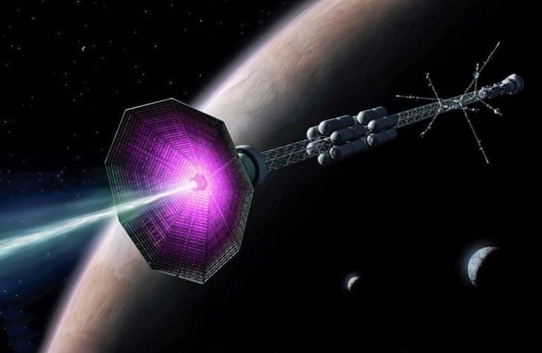
Toggle light / dark theme

Queen’s University researchers uncover brain-based marker of new thoughts and discover we have more than 6,000 thoughts each day.

Researchers at Queen’s University have established a method that, for the first time, can detect indirectly when one thought ends and another begins. Dr. Jordan Poppenk (Psychology) and his master’s student, Julie Tseng, devised a way to isolate “thought worms,” consisting of consecutive moments when a person is focused on the same idea. This research was recently published in Nature Communications.

“What we call thought worms are adjacent points in a simplified representation of activity patterns in the brain. The brain occupies a different point in this ‘state space’ at every moment. When a person moves onto a new thought, they create a new thought worm that we can detect with our methods,” explains Dr. Poppenk, who is the Canada Research Chair in Cognitive Neuroscience. “We also noticed that thought worms emerge right as new events do when people are watching movies. Drilling into this helped us validate the idea that the appearance of a new thought worm corresponds to a thought transition.”

Scientists successfully edited RNA in a living animal in such a way that the repaired RNA then corrected a mutation in a protein that gives rise to a debilitating neurological disorder in people known as Rett syndrome.

The advance by researchers at Oregon Health & Science University publishes in the journal Cell Reports.

“This is the first example of using programmable RNA editing to repair a gene in mouse models of a neurological disease,” said senior author Gail Mandel, Ph.D., senior scientist in the OHSU Vollum Institute. “This gives us an approach that has some traction.”

A new method developed at Cold Spring Harbor Laboratory (CSHL) uses DNA sequencing to efficiently map long-range connections between different regions of the brain. The approach dramatically reduces the cost of mapping brain-wide connections compared to traditional microscopy-based methods.

Neuroscientists need anatomical maps to understand how information flows from one region of the to another. “Charting the cellular connections between different parts of the brain—the connectome—can help reveal how the nervous system processes information, as well as how faulty wiring contributes to and other disorders,” says Longwen Huang, a postdoctoral researcher in CSHL Professor Anthony Zador’s lab. Creating these maps has been expensive and time-consuming, demanding massive efforts that are out of reach for most research teams.

Researchers usually follow neurons’ paths using , which can highlight how individual cells branch through a tangled neural network to find and connect with their targets. But, the palette of fluorescent labels suitable for this work is limited. Researchers can inject different colored dyes into two or three parts of the brain, then trace the connections emanating from those regions. They can repeat this process, targeting new regions, to visualize additional connections. In order to generate a brain-wide map, this must be done hundreds of times, using new research animals each time.

#HubbleClassic On January 19, 2015, Hubble captured a global map of Jupiter. This video was made from the observations.

Today, Jupiter is at opposition, meaning it shines in our sky all night long and is the closest to Earth that it’ll be all year.

#NASA #Hubble #classic #jupiter #planet #video #solarsystem #astronomy #space #science

Great.


NASA is sending a small helicopter called Ingenuity with the Perseverance rover to Mars. It will be the first aircraft to fly in Mars if all goes well. [NASA has built a helicopter to explore Mars and it’s finally ready to launch](https://www.space.com/first-mars-helicopter-ready-to-launch.html)

Credit: NASA/JPL-Caltech

Moderna, Inc.’s COVID-19 vaccine candidate mRNA-1273 will advance to a 30,000-participant Phase III trial later this month, following publication of additional positive Phase I data from a study led by the National Institutes of Health (NIH)’s National Institute of Allergy and Infectious Diseases (NIAID).


Moderna said its closely-watched COVID-19 vaccine candidate mRNA-1273 will advance to a 30,000-participant Phase III trial later this month, following publication of additional positive Phase I data from a study led by the NIH’s National Institute of Allergy and Infectious Diseases (NIAID).

The Phase III “COVE” study (NCT04470427) is expected to begin registration at study centers nationwide beginning on July 21 with study initiation set for six days later. The primary endpoint of the randomized, 1:1 placebo-controlled trial will be the prevention of symptomatic COVID-19 disease. Key secondary endpoints include prevention of severe COVID-19 disease as defined by the need for hospitalization, and prevention of infection by SARS-CoV-2, Moderna said.

Moderna disclosed plans for the Phase III trial on Clinicaltrials.gov the same day that researchers from NIAID, Moderna, and their clinical research partners reported that mRNA-1273 induced rapid and strong immune responses against SARS-CoV-2, in an interim analysis of results from their Phase I study (NCT04283461).

The first episode of “Hubble – Eye in the Sky” is here! 🎥

Have you ever wondered how a telescope in space is operated from down on Earth? Welcome to the Hubble control center. Find out how extraordinarily detailed observations are made with an orbiting space telescope, and get a look at Hubble’s on-the-ground replica that engineers and scientists use down here to fix problems up there.

For more on this video series, visit https://go.nasa.gov/3eveFO1.

Scientists from Trinity College Dublin have discovered a new link between impaired brain energy metabolism and delirium—a disorienting and distressing disorder particularly common in the elderly and one that is currently occurring in a large proportion of patients hospitalized with COVID-19 [15th of July 2020].

While much of the research was conducted in mice, additional work suggests overlapping mechanisms are at play in humans because cerebrospinal fluid (CSF) collected from patients suffering from delirium also contained tell-tale markers of altered brain glucose .

Collectively, the research, which has just been published in the Journal of Neuroscience, suggests that therapies focusing on brain energy metabolism may offer new routes to mitigating delirium.