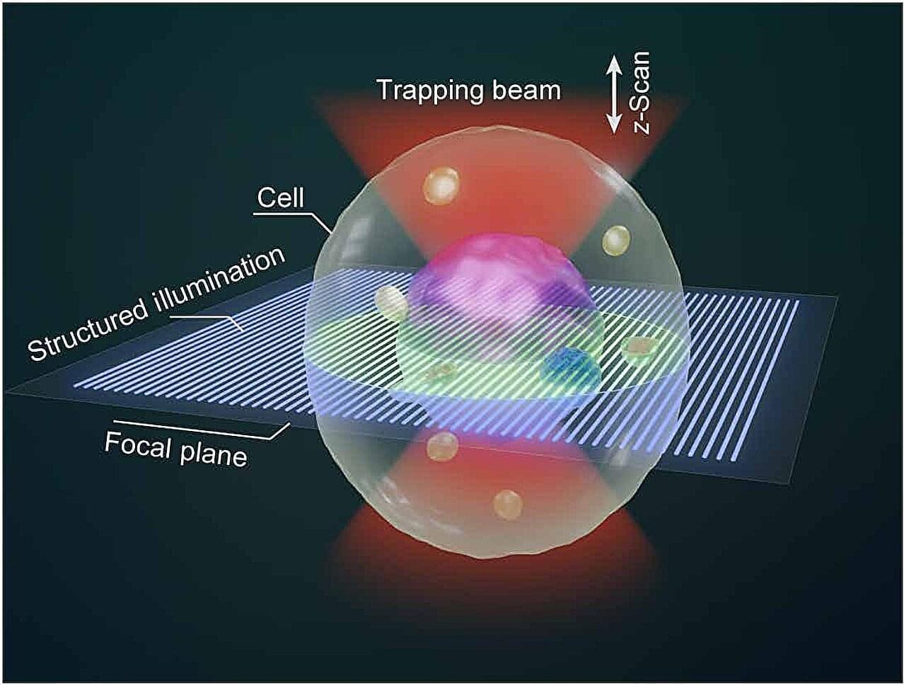
Three-dimensional (3D) imaging is essential for investigating cellular structure and dynamics. Traditional optical methods rely on adhesive or mechanical forces to hold and scan cells, which limit their applicability to suspended cells and may induce stress responses. Developing a non-contact, all-optical 3D imaging technique for live suspended cells remains a major challenge in advancing in situ biological research.
In a study published in Science Advances, Prof. Yao Baoli from the Xi’an Institute of Optics and Precision Mechanics (XIOPM) of the Chinese Academy of Sciences and Prof. Olivier J. F. Martin, from the Swiss Federal Institute of Technology, Lausanne, developed the optical tweezer sectioning microscopy (OTSM), enabling all-optical 3D imaging of suspended live cells, which offers a powerful new tool for live-cell imaging, dynamic biological studies and multicellular assembly.
Researchers developed OTSM by integrating holographic optical tweezers (HOTs) with structured illumination microscopy (SIM).


