Toggle light / dark theme

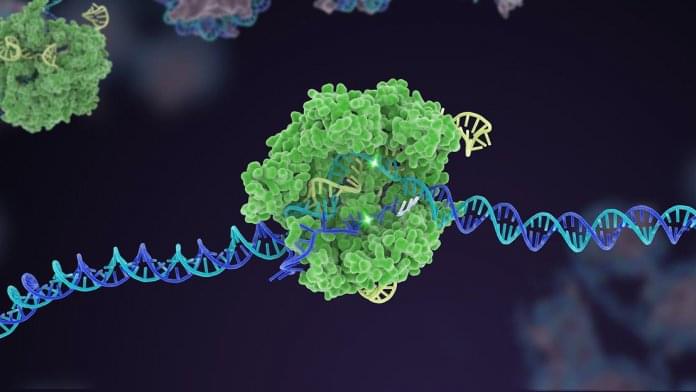
A team of physicists has discovered how DNA molecules self-organize into adhesive patches between particles in response to assembly instructions. Its findings offer a “proof of concept” for an innovative way to produce materials with a well-defined connectivity between the particles.

The work is reported in Proceedings of the National Academy of Sciences.

“We show that one can program particles to make tailored structures with customized properties,” explains Jasna Brujic, a professor in New York University’s Department of Physics and one of the researchers. “While , drills, and hammers must be controlled by humans in constructing buildings, this work reveals how one can use physics to make that ‘know’ how to assemble themselves.”

The device you are currently reading this article on was born from the silicon revolution. To build modern electrical circuits, researchers control silicon’s current-conducting capabilities via doping, which is a process that introduces either negatively charged electrons or positively charged “holes” where electrons used to be. This allows the flow of electricity to be controlled and for silicon involves injecting other atomic elements that can adjust electrons—known as dopants—into its three-dimensional (3D) atomic lattice.

Silicon’s 3D lattice, however, is too big for next-generation electronics, which include ultra-thin transistors, new devices for optical communication, and flexible bio-sensors that can be worn or implanted in the human body. To slim things down, researchers are experimenting with materials no thicker than a single sheet of atoms, such as . But the tried-and-true method for doping 3D silicon doesn’t work with 2D graphene, which consists of a single of carbon atoms that doesn’t normally conduct a current.

Rather than injecting dopants, researchers have tried layering on a “charge-transfer layer” intended to add or pull away electrons from the graphene. However, previous methods used “dirty” materials in their charge-transfer layers; impurities in these would leave the graphene unevenly doped and impede its ability to conduct electricity.

Integrated quantum photonics (IQP) is a promising platform for realizing scalable and practical quantum information processing. Up to now, most of the demonstrations with IQP focus on improving the stability, quality, and complexity of experiments for traditional platforms based on bulk and fiber optical elements. A more demanding question is: “Are there experiments possible with IQP that are impossible with traditional technology?”

This question is answered affirmatively by a team led jointly by Xiao-Song Ma and Labao Zhang from Nanjing University, and Xinlun Cai from Sun Yat-sen University, China. As reported in Advanced Photonics, the team realizes quantum communication using a chip based on silicon photonics with a superconducting nanowire single-photon detector (SNSPD). The excellent performance of this chip allows them to realize optimal time-bin Bell state measurement and to significantly enhance the key rate in quantum communication.

The single photon detector is a key element for quantum key distribution (QKD) and highly desirable for photonic chip integration to realize practical and scalable quantum networks. By harnessing the unique high-speed feature of the optical waveguide-integrated SNSPD, the dead time of single-photon detection is reduced by more than an order of magnitude compared to the traditional normal-incidence SNSPD. This in turn allows the team to resolve one of the long-standing challenges in quantum optics: Optimal Bell-state measurement of time-bin encoded .

Scientists at the John A. Moran Eye Center at the University of Utah have discovered a new type of nerve cell, or neuron, in the retina.

In the central nervous system, a complex circuitry of neurons communicate with each other to relay sensory and motor information; so-called interneurons serve as intermediaries in the chain of communication. Publishing in the Proceedings of the National Academy of Sciences of the United States of America, a research team led by Ning Tian, Ph.D., identifies a previously unknown type of interneuron in the mammalian .

The discovery marks a notable development for the field as scientists work toward a better understanding of the central nervous system by identifying all classes of neurons and their connections.

Two drugs approved decades ago not only counteract brain damage caused by Alzheimer’s disease in animal models, the same therapeutic combination may also improve cognition.

Sounds like a slam dunk in terms of a cure—but not yet. Researchers currently are concentrating on animal studies amid implications that remain explosive: If a surprising drug combination continues to destroy a key feature of the disease, then an effective treatment for Alzheimer’s may have been hiding for decades in plain sight.

A promising series of early studies is highlighting two well known medicine cabinet standbys—gemfibrosil, an old-school cholesterol-lowering drug, and retinoic acid, a vitamin A derivative. Gemfibrosil, is sold as Lopid and while it’s still used, it is not widely prescribed. Doctors now prefer to prescribe statins to lower cholesterol. Retinoic acid has been used in various formulations to treat everything from acne to psoriasis to cancer.

On a chilly evening last fall, I stared into nothingness out of the floor-to-ceiling windows in my office on the outskirts of Harvard’s campus. As a purplish-red sun set, I sat brooding over my dataset on rat brains. I thought of the cold windowless rooms in downtown Boston, home to Harvard’s high-performance computing center, where computer servers were holding on to a precious 48 terabytes of my data. I have recorded the 13 trillion numbers in this dataset as part of my Ph.D. experiments, asking how the visual parts of the rat brain respond to movement.

Printed on paper, the dataset would fill 116 billion pages, double-spaced. When I recently finished writing the story of my data, the magnum opus fit on fewer than two dozen printed pages. Performing the experiments turned out to be the easy part. I had spent the last year agonizing over the data, observing and asking questions. The answers left out large chunks that did not pertain to the questions, like a map leaves out irrelevant details of a territory.

But, as massive as my dataset sounds, it represents just a tiny chunk of a dataset taken from the whole brain. And the questions it asks—Do neurons in the visual cortex do anything when an animal can’t see? What happens when inputs to the visual cortex from other brain regions are shut off?—are small compared to the ultimate question in neuroscience: How does the brain work?

None.


Scientists from Heidelberg and Bern have succeeded in training spiking neural networks to solve complex tasks with extreme energy efficiency. The advance was enabled by the BrainScaleS-2 neuromorphic platform, which can be accessed online as part of the EBRAINS research infrastructure.

Developing a machine that processes information as efficiently as the human brain has been a long-standing research goal towards true artificial intelligence. An interdisciplinary research team at Heidelberg University and the University of Bern led by Dr Mihai Petrovici is tackling this problem with the help of biologically-inspired artificial neural networks.

Spiking neural networks, which mimic the structure and function of a natural nervous system, represent promising candidates because they are powerful, fast, and energy-efficient. One key challenge is how to train such complex systems. The German-Swiss research team has now developed and successfully implemented an algorithm that achieves such training.