Toggle light / dark theme

In a study recently published in the journal Nano Letters, researchers from Nano Life Science Institute (WPI-NanoLSI), Kanazawa University, Kanazawa, Japan, used frequency-modulated atomic force microscopy to reveal the submolecular structure of microtubule (MT) inner surface and visualize structural defects in the MT lattice, providing valuable insights into the complex dynamic processes that regulate microtubule function.

Microtubules (MTs), a key component of the cytoskeleton in , serve as scaffolds and play vital roles in cellular processes such as , cell migration, intracellular transport, and trafficking. MTs are composed of α-tubulin and β-tubulin proteins, which polymerize into dimers and assemble into linear protofilaments that form a cylindrical lattice.

Traditional methods like X-ray crystallography and cryo– have provided structural insights into MTs but involve complex sample preparation and data analysis. There remains a need for techniques that can examine MT structural features, assembly dynamics, and lattice defects at submolecular resolution under physiological conditions.

At just a few atoms of thickness, 2D materials offer revolutionary possibilities for new technologies that are microscopically sized but have the same capabilities as existing machines.

Florida State University researchers have unlocked a new method for producing one class of 2D material and for supercharging its magnetic properties. The work was published in Angewandte Chemie.

Experimenting on a metallic magnet made from the elements iron, germanium and tellurium and known as FGT, the research team made two breakthroughs: a collection method that yielded 1,000 times more material than typical practices, and the ability to alter FGT’s magnetic properties through a chemical treatment.

Here the authors report PICNIC (Proteins Involved in CoNdensates In Cells), a machine learning algorithm that predicts approximately 40–60% of proteins form condensates in various organisms, showing no clear relationship with the complexity of the organism or the content of disordered proteins.

Google’s new quantum computing chip, Willow, has set a groundbreaking standard by achieving unparalleled speed and precision, outperforming supercomputers in specific tasks by millions of times. This revolutionary chip enhances quantum error correction, making scalable quantum systems a reality and unlocking new possibilities for artificial intelligence, scientific research, and real-world problem-solving. Willow’s success marks a major milestone in the integration of quantum computing and AI, driving innovation across industries.

Don’t let AI leave you behind — get updates at https://airevolution.cc.

🔍 Key Topics:
Google’s Willow chip and its revolutionary quantum computing advancements.
How quantum error correction enables scalable and stable systems with unmatched performance.
The integration of quantum computing and AI to tackle problems beyond classical limits.

🎥 What You’ll Learn:
Why Willow represents a major breakthrough in quantum computing and AI innovation.
How it reduces errors and enhances performance for real-world applications in medicine, energy, and more.
The potential of quantum AI to transform industries and solve previously unsolvable challenges.

📊 Why It Matters:
This video explores Google’s revolutionary quantum chip, Willow, and its impact on the future of computation and AI, highlighting its scalability, precision, and groundbreaking applications in science and industry.

DISCLAIMER:

The preprint, not yet peer-reviewed, is the latest from a global consortium that hunts down potential new genes. Ever since the Human Genome Project completed its first draft at the turn of the century, scientists have tried to decipher the genetic book of life. Buried within the four genetic letters—A, T, C, and G—and the proteins they encode is a wealth of information that could help tackle our most frustrating medical foes, such as cancer.

The Human Genome Project’s initial findings came as a surprise. Scientists found less than 30,000 genes that build our bodies and keep them running—roughly a third of that previously predicted. Now, roughly 20 years later, as the technologies that sequence our DNA or map proteins have become increasingly sophisticated, scientists are asking: “What have we missed?”

The new study filled the gap by digging into relatively unexplored portions of the genome. Called “non-coding,” these parts haven’t yet been linked to any proteins. Combining several existing datasets, the team zeroed in on thousands of potential new genes that make roughly 3,000 miniproteins.

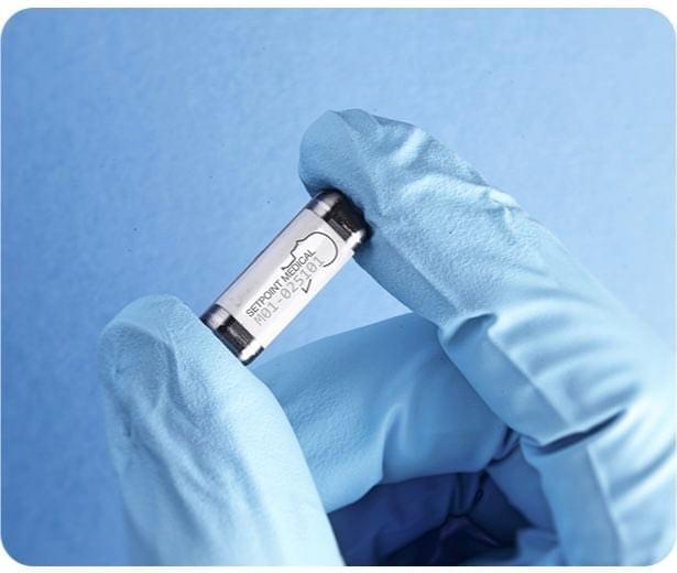
Tiny implantable sensors are helping University of Oregon researchers optimize the process of recovery from severe bone injuries.

Scientists at the UO’s Phil and Penny Knight Campus for Accelerating Scientific Impact have developed miniature that transmit about what’s happening at an injury site. In a new study, they use the technology to show that a resistance-training rehabilitation program can significantly improve femur injuries in rats in just eight weeks.

The sensors provide a window into the mechanical properties of the , giving scientists detailed ongoing data about the process of . If someday applied in humans, these sensors could allow doctors to better tailor a rehabilitation program to an individual patient, monitoring their progress and adjusting the exercises along the way.