Synaptic Density in Multiple Sclerosis: An In Vivo Study Using [11C]UCB-J-PET Imaging.
Background and Objectives.


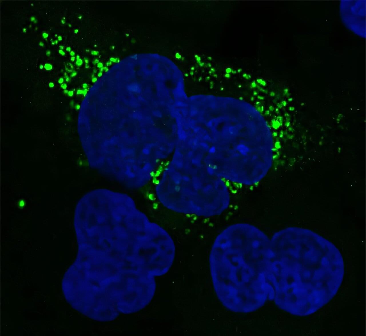
This review explores the current literature on brain MRI findings of CAR-T–induced neurotoxicity, highlighting diagnostic capabilities, clinical implications, and emerging trends in advancing imaging modalities.
Chimeric antigen receptor T-cell (CAR-T) therapy has remarkable efficacy in treating refractory hematologic malignancies. However, CAR-T therapy may induce neurotoxic effects in some patients. Common symptoms of neurotoxicity range from early signs such as headache, confusion, delirium, and aphasia to severe manifestations such as seizures, motor weakness, increased intracranial pressure, cerebral edema, and coma. Magnetic resonance imaging (MRI) can offer invaluable insight into resulting abnormalities in the structure, physiology, and function of the central nervous system. This review aims to examine the current literature on brain MRI findings of CAR-T–induced neurotoxicity, elucidating its diagnostic capabilities, clinical implications, and emerging trends in advancing imaging modalities.

Silicon photonics is a revolutionary technology in the integrated photonics field which has experienced rapid development over the past several decades. High-quality III-V semiconductor components on Si platforms have shown their great potential to realize on-chip light-emitting sources for Si photonics with low-cost and high-density integration. In this review, we will focus on semiconductor optical amplifiers (SOAs), which have received considerable interest in diverse photonic applications. SOAs have demonstrated high performance in various on-chip optical applications through different integration technologies on Si substrates. Moreover, SOAs are also considered as promising candidates for future light sources in the wavelength tunable laser, which is one of the key suitable components in coherent optical devices.
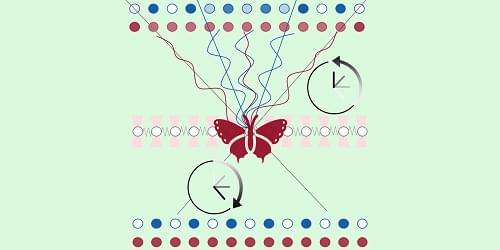

Light can behave in very unexpected ways when you squeeze it into small spaces. In a paper in the journal Science, Mark Brongersma, a professor of materials science and engineering at Stanford University, and doctoral candidate Skyler Selvin describe the novel way they have used sound to manipulate light that has been confined to gaps only a few nanometers across—allowing the researchers exquisite control over the color and intensity of light mechanically.
The findings could have broad implications in fields ranging from computer and virtual reality displays to 3D holographic imagery, optical communications, and even new ultrafast, light-based neural networks.
The new device is not the first to manipulate light with sound, but it is smaller and potentially more practical and powerful than conventional methods. From an engineering standpoint, acoustic waves are attractive because they can vibrate very fast, billions of times per second.
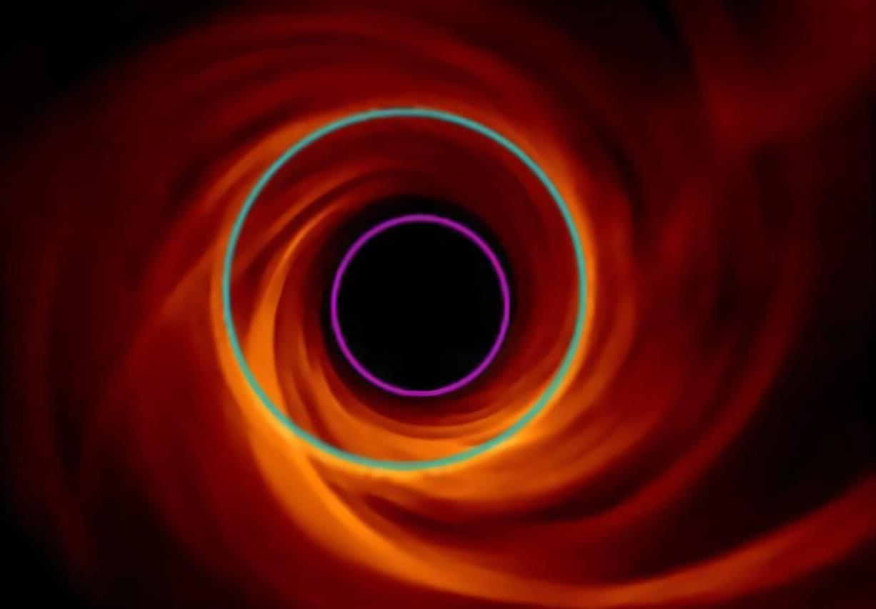
The first black hole images stunned the world in 2019, with headlines announcing evidence of a glowing doughnut-shaped object from the center of galaxy Messier 87 (M87 —55 million light years from Earth. Supercomputer simulations are now helping scientists sharpen their understanding about the environment beyond a black hole’s ‘shadow,’ material just outside its event horizon.
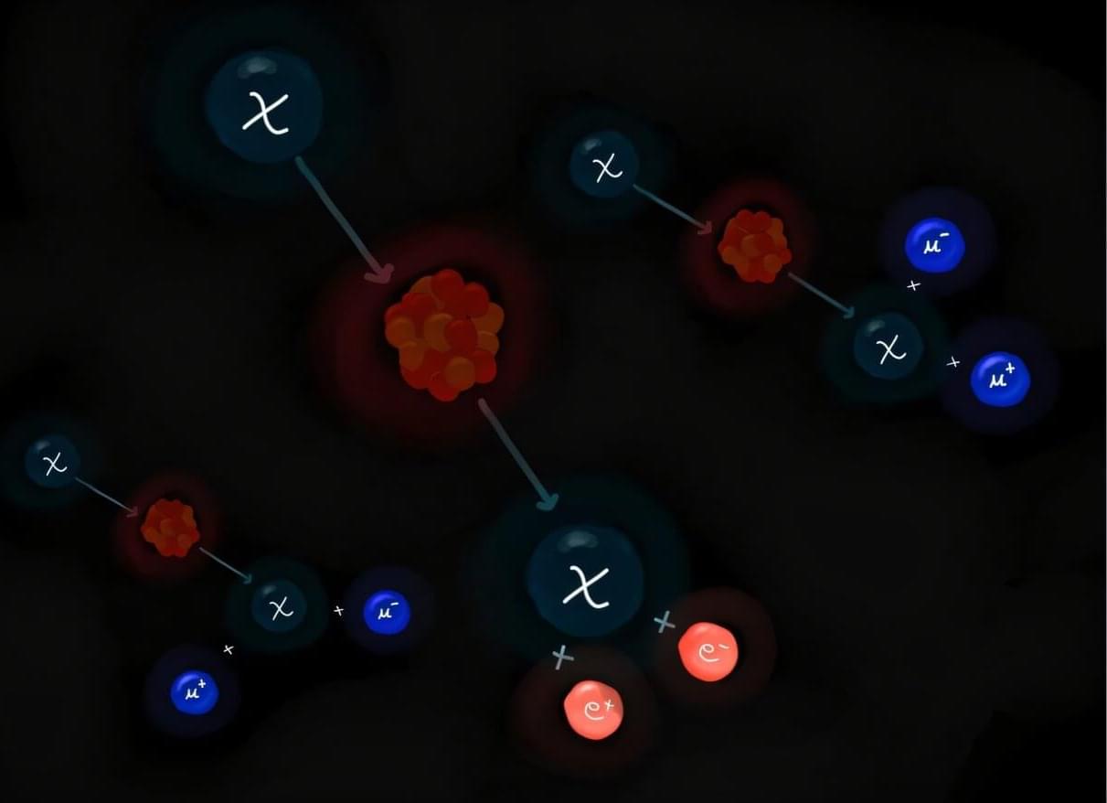
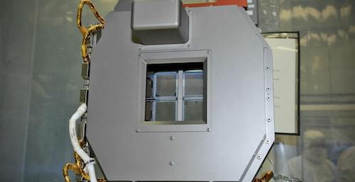
Dark matter (DM) is a type of matter estimated to account for 80% of the universe’s total mass, but it cannot be directly detected using conventional experimental techniques. As DM does not emit, reflect or absorb light, most previous dark matter searches were aimed at observing either its weak interactions with ordinary matter using highly sensitive detectors or other signatures linked to its presence or decay.
Researchers at Texas A&M University recently introduced a new approach that could enable the direct detection of this elusive type of matter, leveraging a process known as the DM internal pair production. Their proposed strategy, outlined in a paper published in Physical Review Letters, could open new possibilities for future DM searches focusing on a wide range of candidate particles.
“The particle nature of DM can be revealed when a DM particle scatters off a nucleus and produces a visible recoil signal,” the authors told Phys.org. “However, for light DM, transferring sufficient energy to a heavy nucleus is kinematically challenging, even if the DM is energetic. To overcome this limitation, we developed a framework where additional particles are produced in the final state, allowing the DM’s energy to be shared among them, while the nucleus remains largely at rest.”

Salt creeping, a phenomenon that occurs in both natural and industrial processes, describes the collection and migration of salt crystals from evaporating solutions onto surfaces. Once they start collecting, the crystals climb, spreading away from the solution. This creeping behavior, according to researchers, can cause damage or be harnessed for good, depending on the context.
New research published June 30 in the journal Langmuir is the first to show salt creeping at a single-crystal scale and beneath a liquid’s meniscus.
“The work not only explains how salt creeping begins, but why it begins and when it does,” says Joseph Phelim Mooney, a postdoc in the MIT Device Research Laboratory and one of the authors of the new study. “We hope this level of insight helps others, whether they’re tackling water scarcity, preserving ancient murals, or designing longer-lasting infrastructure.”