As nearly one in six couples experience fertility issues, in-vitro fertilization (IVF) is an increasingly common form of reproductive technology. However, there are still many unanswered scientific questions about the basic biology of embryos, including the factors determining their viability, that, if resolved, could ultimately improve IVF’s success rate.
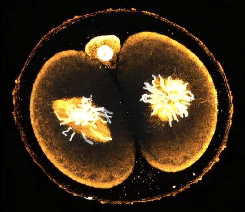
A new study from Caltech examines mouse embryos when they are composed of just two cells, right after undergoing their very first cellular division. This research is the first to show that these two cells differ significantly—with each having distinct levels of certain proteins.
Importantly, the research reveals that the cell that retains the site of sperm entry after division will ultimately make up the majority of the developing body, while the other largely contributes to the placenta. While the studies were done in mouse models, they provide critical direction for understanding how human embryos develop. Indeed, the researchers also assessed human embryos immediately after their first cellular division and found that these two cells are likewise profoundly different.