Toggle light / dark theme

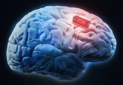
(Medical Xpress)—A team of researchers working at the University of Rochester in New York, has found that injecting glial cells into a mouse brain caused an improvement in both memory and cognition in the mouse. In their paper published in The Journal of Neuroscience, the team explains how they injected the test mice and then tested them afterwards to see what impact it had on their abilities.

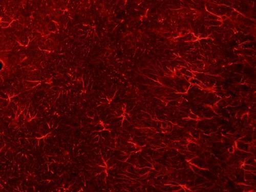
Injecting human into mice brains appears to be the stuff of horror films, but in this case, it wasn’t really what it might have seemed. Glial cells are precursors to other cells—in this case, they develop into astrocytes, which are technically, brain cells. But, the important distinction here is that they are not neurons, which means they are not involved in thinking—instead they are involved in memory retention and help with housekeeping tasks.

Last year, the team injected mature into mice brains and reported improvements in ability by the mice—this time they went further, injecting progenitor precursor glial cells, which allows for development of more astrocytes. The team injected just 300,000 of the cells (from donated human embryos) and found just 12 months later that they had multiplied to grow to 12 million, completely displacing the original mouse astrocytes. It appeared, the team reported, that the cell growth only stopped when it reached the physical confines of the skull. They also note that it was interesting that the glial could thrive in such an environment considering that astrocytess in people are 10 to 20 times as big as those in mice and they carry 100 times as many tendrils. Testing the mice showed that their memory was far superior to normal mice and they had improved cognition as well.
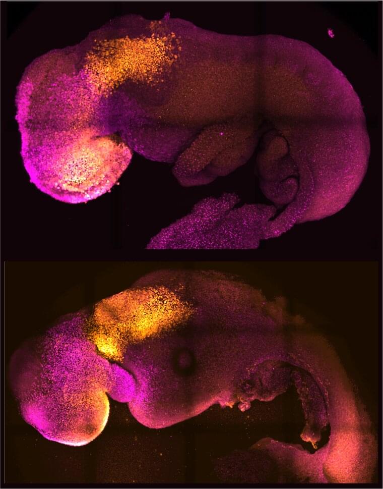
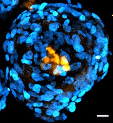
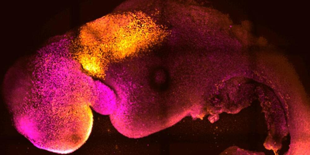
Researchers from the University of Cambridge and Caltech have created model mouse embryos from stem cells—the body’s master cells, which can develop into almost any cell type in the body—that have beating hearts, as well as the foundations for a brain and all of the other organs in the mouse body.

The results are the culmination of more than a decade of research, and they could help researchers understand why some embryos fail while others go on to develop into a fetus as part of a healthy pregnancy. Additionally, the results could be used to guide repair and development of synthetic human organs for transplantation.

The research was conducted in the laboratory of Magdalena Zernicka-Goetz, Bren Professor of Biology and Biological Engineering at Caltech. Zernicka-Goetz is also a professor of mammalian development and in Cambridge’s Department of Physiology, Development and Neuroscience. A paper describing the breakthrough appears in the journal Nature on August 25.

Research on human embryos is vital to understanding the earliest stages of human development. Currently, this research is conducted on surplus embryos willingly donated by individuals who have undergone in vitro fertilization. Nevertheless, this research is limited by the availability of embryos and strict international ethical time limits on how long an embryo is allowed to develop in the laboratory (14 days maximum.)

Now, Caltech researchers have created embryo-like structures out of human stem cells. In contrast to natural embryos that are formed by a combination of sperm and egg, these structures are formed by combining so-called , which have the ability to develop into specialized types of cells. Though these embryo-like structures have some key differences from real embryos, the technology to create them will be critical in answering open questions about without the need for donated embryos.

The research was conducted in the laboratory of Magdalena Zernicka-Goetz, Bren Professor of Biology and Biological Engineering at Caltech, and is described in a paper appearing in the journal Nature Communications on September 21.

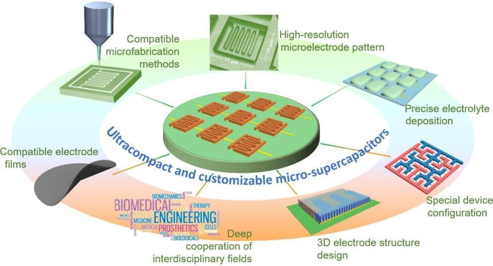
Increased demand for super tiny electronic sensors coming from healthcare, environmental services and the Internet of Things is prompting a search for equally tiny ways to power these sensors. A review of the state of ultracompact supercapacitors, or “micro-supercapacitors,” concludes there is still a lot of research to be done before these devices can deliver on their promise.

The review appeared in the journal Nano Research Energy.

The explosion of demand in recent years for miniaturized , such as health monitors, environmental sensors and wireless communications technologies has in turn driven demand for components for those devices that have ever smaller size and weight, with lower energy consumption, and all of this at cheaper prices.

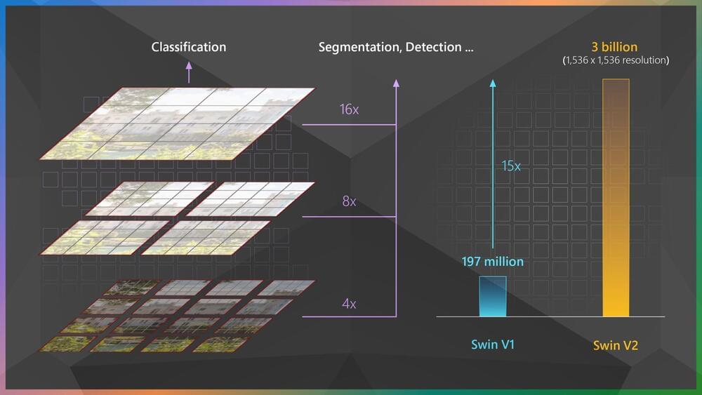
Early last year, our research team from the Visual Computing Group introduced Swin Transformer, a Transformer-based general-purpose computer vision architecture that for the first time beat convolutional neural networks on the important vision benchmark of COCO object detection and did so by a large margin. Convolutional neural networks (CNNs) have long been the architecture of choice for classifying images and detecting objects within them, among other key computer vision tasks. Swin Transformer offers an alternative. Leveraging the Transformer architecture’s adaptive computing capability, Swin can achieve higher accuracy. More importantly, Swin Transformer provides an opportunity to unify the architectures in computer vision and natural language processing (NLP), where the Transformer has been the dominant architecture for years and has benefited the field because of its ability to be scaled up.

So far, Swin Transformer has shown early signs of its potential as a strong backbone architecture for a variety of computer vision problems, powering the top entries of many important vision benchmarks such as COCO object detection, ADE20K semantic segmentation, and CelebA-HQ image generation. It has also been well-received by the computer vision research community, garnering the Marr Prize for best paper at the 2021 International Conference on Computer Vision (ICCV). Together with works such as CSWin, Focal Transformer, and CvT, also from teams within Microsoft, Swin is helping to demonstrate the Transformer architecture as a viable option for many vision challenges. However, we believe there’s much work ahead, and we’re on an adventurous journey to explore the full potential of Swin Transformer.

In the past few years, one of the most important discoveries in the field of NLP has been that scaling up model capacity can continually push the state of the art for various NLP tasks, and the larger the model, the better its ability to adapt to new tasks with very little or no training data. Can the same be achieved in computer vision, and if so, how?

Astronomers build new telescopes and peer at the night sky to see what they might find. Janelia Group Leader Abraham Beyene takes a similar approach when looking at the cells that make up the human brain.

Beyene and his team design and synthesize new types of highly sensitive biosensors they use to peer at to see what they can learn.

“You have this new tool that now helps us make the kinds of measurements that we’ve never been able to make before, and we go into the lab and deploy this technology and we see what happens,” Beyene says. “What you see is that some really interesting phenomena begin to emerge that you haven’t even begun to think about.”

Scientists have created “synthetic” mouse embryos from stem cells without a dad’s sperm or a mom’s egg or womb.

The lab-created embryos mirror a natural mouse embryo up to 8 ½ days after fertilization, containing the same structures, including one like a beating heart.

In the near term, researchers hope to use these so-called embryoids to better understand early stages of development and study mechanisms behind disease without the need for as many lab animals. The feat could also lay the foundation for creating synthetic human embryos for research in the future.